Year: 2022

20 May 2022
Orthopantomography of an adult patient, dentistry

X-Rays and Radiation Safety Principles in Dentistry

Dental x-rays are important for maintaining an individual’s optimal oral health. Like with any x-ray procedure, there are radiation safety principles that have been put in place to protect both patients and dentistry professionals.

Why are Dental X-rays Important?

Dental x-rays are an essential part of maintaining a healthy jaw and teeth. This diagnostic tool allows dentists to take a more in-depth look at what’s going on inside a patient’s mouth. Some dental problems affecting the roots of a tooth or that exist below the gum line cannot be diagnosed through a simple visual exam.

What Can a Dental X-ray Be Used for?

Dentists use dental x-rays to diagnose a variety of dental conditions, including:

  • Gum disease
  • Cavities and tooth decay
  • Impacted teeth
  • Cysts
  • Abscesses
  • Jaw disorders
  • Sinus issues
  • Bone loss

These x-rays are also beneficial for individuals who smoke, have a history of restorative dental work, or drink an excessive number of sugary drinks like pop or juice.

Radiation Safety for Patients During Dental X-rays

Most patients will undergo at least one of two different types of dental x-rays during their annual or bi-annual visit to the dentist.  Each has its own radiation protection requirements to limit the patient’s radiation dose.

Bitewing X-rays

Bitewing x-rays are a type of intraoral x-ray. During a bitewing x-ray, the patient is required to bite down on a small tab attached to an x-ray film. There are usually 4 images taken to help dentists visualize all sides of the mouth.

Patients are usually required to wear a lead vest during a bitewing x-ray. Thyroid collars and leaded glasses can also be used to protect the eyes and sensitive areas of the throat.
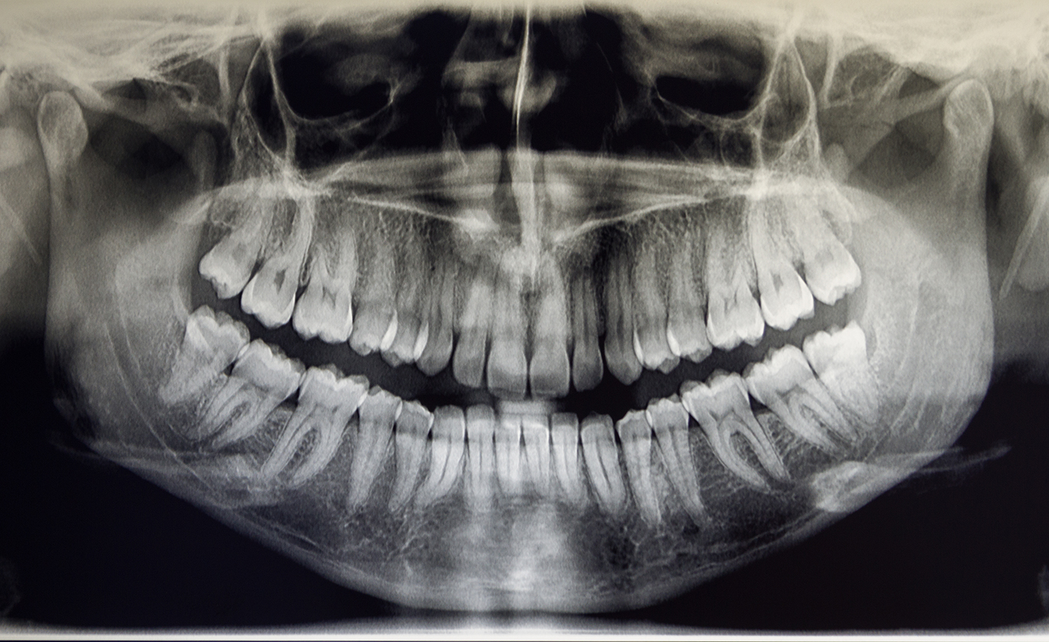
Panoramic X-rays

Panoramic x-rays are extraoral x-rays used to diagnose dental problems in the jaw and skull. This 2D digital x-ray captures a single image of the entire mouth.

dental x-ray

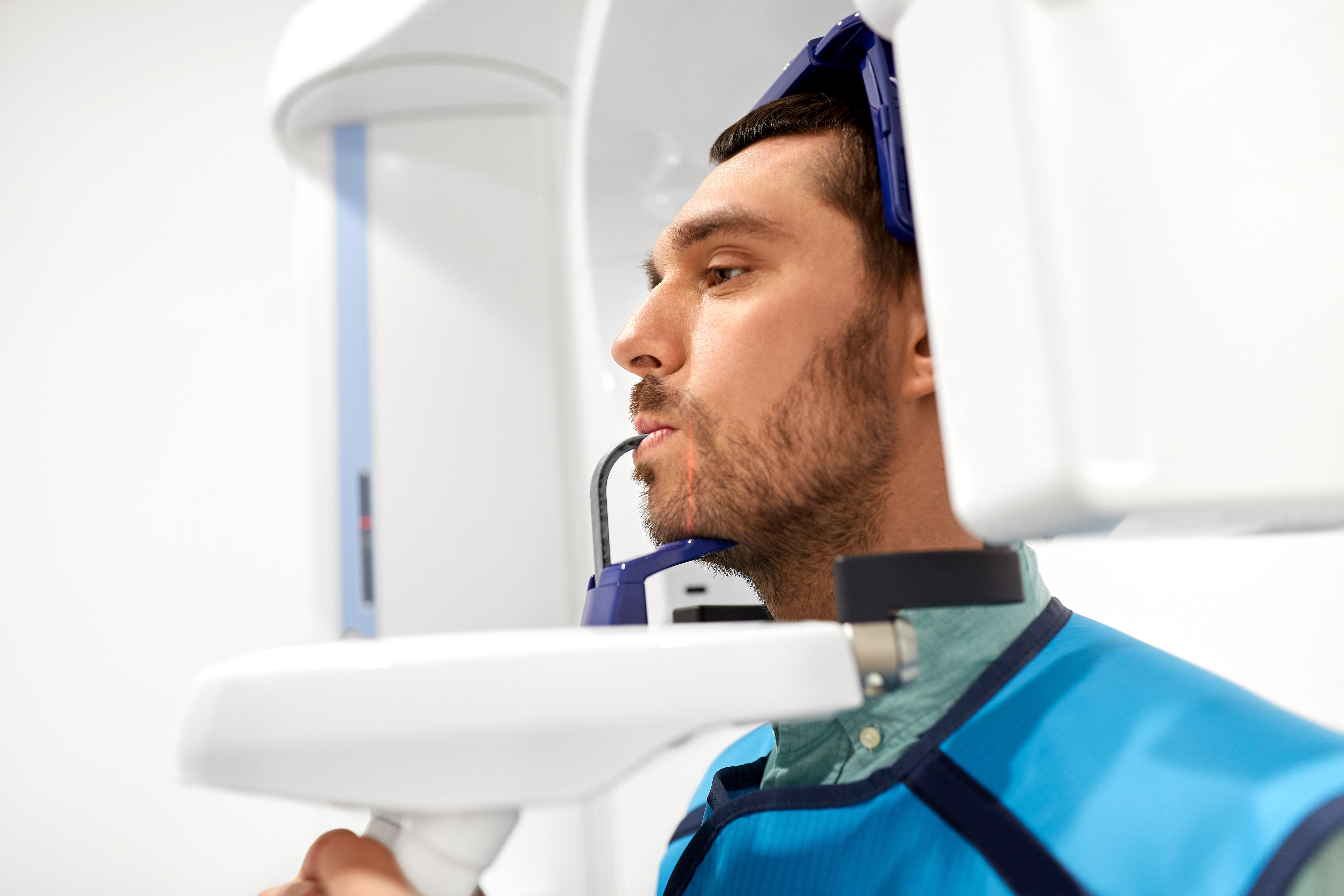
The machine has two sides. One side houses the x-ray tube, and the other houses the x-ray detector or x-ray film. The x-ray rotates around the patient’s head during this procedure to take the full image. A lead apron or thyroid protective collar may also be worn during this procedure as a safety precaution; however, it is not required.

Miscellaneous Dental X-rays

Other types of dental x-rays a patient may experience include a dental cone beam CT scan (3D image of the teeth and jaw) or a cephalometric projection (image of the entire head). These x-rays are not likely to occur during a routine visit.

Are Dental X-rays Safe?

Patients are exposed to extremely minimal radiation doses during dental x-ray procedures. A patient who undergoes a bitewing x-ray procedure will receive a radiation dose of 0.4 mrem, while a panoramic x-ray radiation dose exposure is approximately 0.7 mrem.  

For comparison, the average U.S. citizen receives an annual radiation dose of 620 mrem. The sources of this radiation come from naturally occurring cosmic, internal, and terrestrial radiation, medical procedures like CT scans and conventional radiography procedures, and consumer products like tobacco, fertilizer, or welding rods.  

As mentioned above, there are numerous protective measures put in place to help limit overall radiation exposure. Protective measures for patients during a dental x-ray include:

  • Using protective lead aprons and thyroid collars
  • Using modern x-ray equipment and up to date imaging techniques
  • Limiting the number of images taken in a year

The American Dental Association recommends that healthy patients receive x-rays once every 2 to 3 years to limit the amount of radiation exposure they receive.

Like any diagnostic procedure, the benefits of a dental x-ray far outweigh the low radiation dose a patient will receive. Dental x-rays can not only identify existing issues, but they can also identify potential problems that, if left untreated, could turn into serious or even life-threatening issues.

Radiation Safety for Dental Professionals

Radiation safety and radiation protection guidelines are especially important for dental professionals. The NRC regulates the amount of radiation exposure for adults who work with radioactive materials. Occupational exposure limits are limited to 5,000 mrem per year.

Dental professionals including hygienists, oral surgeons, and orthodontists, are required to adhere to these exposure limits. This profession receives less ionizing radiation exposure than other healthcare professionals on average. However, basic protective measures help limit their occupational radiation exposure and keep up with ALARA standards.

Personal Dosimeter

Dental professionals should wear a personal dosimeter when operating x-ray equipment to record their dose.

Instadose+ Dosimeter

The Instadose+ dosimeter is an affordable personal dosimeter option that provides dentists and dental professionals with accurate, long-term exposure tracking. The device wirelessly captures, transmits, measures, and analyzes radiation dose exposure. Dental professionals can access their read history on-demand and in real-time thanks to the device’s Bluetooth and SmartMonitoring technology.

Unlike traditional film badges, the Instadose+ dosimeter does not need to be collected and processed off-site.

Time, Distance, and Shielding

The standard guidelines for minimizing dental professionals’ exposure are time, distance, and shielding. This means limiting the amount of time spent around a radiation source, maintaining a safe distance away from the source of radiation, and standing behind protective barriers during a procedure.

During a routine dental visit, hygienists are often the ones who prepare and take the dental x-ray. To limit their exposure from taking these images multiple times a day, hygienists will usually leave the room to increase their distance from the x-ray tube. They may also stand behind a lead wall or partition while the image is being taken.

The Takeaway

Dental x-rays are a necessary part of maintaining good oral health. Radiation safety guidelines like wearing lead vests and thyroid collars are important for limiting patient exposure during routine checkups. Wearing a personal dosimeter and following time, distance, and shielding guidelines are also necessary for limiting occupational exposure.

28 Apr 2022
Radiation Protection Survey of Package with Pancake Probe

A Beginner’s Guide to Radiation Protection Surveys

The purpose of a radiation protection survey is to identify higher-than-normal doses of radiation in medical environments, labs, and anywhere radiation-emitting machines or radioactive materials (RAM) are used. They are required by state and federal regulations to be performed regularly to ensure the safety of technicians, technologists, nurses, doctors, researchers, and patients.

In this brief guide we’ll talk about what a radiation protection survey is, why it is important, and the type of equipment required to perform a radiation protection survey.

What is a radiation protection survey?

Radiation protection surveys are a way to directly measure radiation levels and identify potential leakage through breaks or voids in shielding.

Surveys are performed on:

  • Diagnostic fluoroscopic and radiographic equipment
  • Non-medical industrial equipment such as those found in veterinary offices
  • CT and CBCT machines
  • Particle accelerators
  • Irradiators
  • Bone mineral densitometers
  • Cabinet x-ray machines
  • Areas that use sealed sources of RAM
  • Packages containing RAM

The Different Types of Radiation Surveys

Not all radiation surveys are created equal. Let’s talk about some of the different surveys you may encounter a need for in your radiation safety program.

Radiation Emitting Device Survey

X-ray machines and other radiation-emitting devices require regular surveys to be performed to confirm that the machine is operating as expected. Radiation producing machines are surveyed for:

  • Timer accuracy
  • Radiation output
  • Focal spot size
  • kVp and mA
  • Beam limitation accuracy
  • Filtration
  • Skin entrance exposure / rate of exposure
  • Scatter radiation measurements
  • Photo-timer operation
  • Proper signage, labels, and postings

If high or unexpected dose rates are measured during a survey, the machine should be turned off and undergo appropriate maintenance.

Area Survey

Area surveys are required anywhere a radiation device is in use and the potential for receiving a higher-than-normal radiation dose is present. These surveys are typically measured in milliRoentgen per hour (mR/hr). The Roentgen is a measure of the amount of ionization in the air from the radiation.

Anytime you have an area survey performed, you are required to keep the official records of the survey results for 3 years.

Contamination Wipe Test

A contamination wipe test, also known as an indirect or swipe survey, is used to identify radioactive material contamination on surfaces, equipment, and clothing such as those found in a lab. This type of survey can identify non-fixed radiation left behind from radioactive solids, liquids, or gasses.

Lab tech performing a wipe test

Wipe tests are recommended to be performed frequently, especially if you are a HAZMAT employee that receives or ships RAM packages. A wipe test involves wiping at least 300cm2 of the package’s surfaces using an absorbent material. Afterward, the activity on the swipe is measured assuming a removal efficiency of 0.1 unless the actual efficiency is known.

Users in lab settings typically survey their work areas after an experiment or when a minor spill is suspected.

Radioactive Sealed Source

A radioactive sealed source is a source of special form RAM that has been contained or encapsulated to prevent contamination. These sources can only be opened by destruction. Semi-annual surveys of these sources are required to check for leakage.

Bioassay Survey

Internal exposure monitoring, or a bioassay survey, is performed on individuals that use unsealed radioactive materials. The survey estimates the internal organ dose to determine if any RAM has entered the body. It can also help determine if RAM is present in the air.

Bioassay surveys are performed by analyzing blood, tissue, or urine samples or by carefully monitoring the presence and/or quality of isotopes present in the organ of concern.

How often do I need to have a survey performed?

The frequency of a radiation protection survey depends on several factors, most of which depend on different state and federal regulations.

  • When a new or used x-ray equipment is installed
  • When existing x-ray equipment has been moved
  • If shielding has been modified
  • After the equipment has undergone significant repairs
  • If a potential problem is indicated

Who performs these surveys?

In general, surveys on radiation-producing equipment are conducted by health physicists and medical physicists.

Is special equipment required for a survey?

Special equipment is required to detect ionizing radiation. Most equipment is hand-held measurement instruments called survey meters. This equipment is required to be calibrated annually to maintain accuracy and to ensure that reliable measurements are recorded.

Survey meters consist of:

  • A probe which produces electrical signals when it is exposed to radiation
  • A control panel readout with an electronic meter that gauges the amount of radiation exposure
  • A speaker which provides an audible indication of the radiation exposure

There are several different kinds of survey meters physicists use to perform radiation surveys.

Geiger-Mueller Pancake Probe

One of the more commonly used survey meters is the Geiger-Mueller Pancake Detector. Although there is no “universal” radiation detector, the G-M Pancake Probe comes pretty close. This is because the probe can detect alpha, beta, and gamma radiation, although they are generally used for detecting Beta Emitters. These probes come in a variety of models and configurations.

Surveying open package with pancake probe

The probe detects radiation by collecting counting gas within the tube. The counting gas is ionized when a photon or particle interacts with a released electron. When the voltage is high, radiation that interacts with the counting gas produces an electronic pulse that is measured with a separate counting instrument.

A pancake probe has a thin layer of mica on the active face of the detector, which allows most alpha and beta particles to interact with the counting gas inside the tube.

G-M Pancake Probes are frequently used to detect C-14, Ca-45, P-32, P-33, and S-35.

Scintillation Survey Meter

A scintillation survey meter is used to detect low-energy Gamma Emitters and x-rays. The scintillator, or sensor, is made of a transparent crystal or liquid which shines when it interacts with ionizing radiation. The scintillator is attached to a photosensor like a photomultiplier tube which detects the generated light.

This survey meter detects I-125 and Cr-51. They are an ideal equipment choice for surveying electron microscopes and x-ray diffractometers.

Diagnostic Physics Support and Radiation Surveys by Versant Physics

When it comes to hiring a consultant to perform radiation protection or QA surveys for your equipment, you want to make sure you’re working with the best. People who are experts in state and federal regulations regarding radiation machines and RAM, have access to top-of-the-line survey equipment and understand the importance of adhering to ALARA standards.

Versant Physics’ proactive and transparent diagnostic physics support process minimizes safety concerns and reduces the likelihood of compliance violations. We support our clients by sharing our knowledge of best practices in advanced technologies, and by utilizing a team-based approach we feel enables our clients to focus on maximizing the quality of patient care.

Contact our team for a free 30-minute consultation to learn more about our diagnostic physics and radiation survey expertise.


20 Apr 2022
nurse guiding patient entering mri scanner

The Basics of Radiation Shielding in Medicine

Basic radiation protection guidelines can be summed up in three simple concepts: time, distance, and shielding. While both limiting the time spent and increasing the proximity to an ionizing radiation source is something that lies within the power of the individual, shielding and X-ray room design require careful planning and execution by the facility or Radiation Safety Officer.

What is radiation shielding?

Radiation shielding is simply a barrier placed between a source of radiation and the area or person that needs to be protected. The purpose of radiation shielding is to limit, control, or modify the radiation exposure rate at a set point.

Shielding is based on attenuation or the gradual reduction in the intensity of energy through a specific medium. X-ray radiation that passes through certain materials decrease and are absorbed, thereby reducing the exposure to the other side of the barrier.

Without shielding, the public, radiation workers (including dentists and veterinarians), and even nearby office workers could be exposed to levels of radiation outside regulated exposure limits, which can potentially lead to negative health effects. Although it is impossible to completely avoid exposure to radiation, shielding is a critical consideration in any medical facility that greatly reduces unnecessary exposure.

Shielding Materials

There are several different materials that provide protection from penetrating radiation. Concrete, water, special plastic shields, air stops, and lead are all barriers that stop different types of rays and particles, reducing the overall dose a person receives.

In medical environments, the most common shielding materials used include lead, lead-free shielding, and lead composites.

Lead

Lead is one of the most used and most effective shielding materials. It is a highly dense material with a high atomic number and a high number of electrons which make it ideal for shielding in most medical radiation environments. This is because the type and energy of radiation in a medical environment that passes through lead are absorbed or scattered by the electrons present in the material.

Vet team wearing shielding garments during exam

Lead is also cheap and easy to process. It can be mixed with other materials like glass, or binders like vinyl, which allows it to be used as construction materials in X-ray rooms or worn as shielding garments.

Lead-Free Shielding

Technological advances have allowed for the creation of non-toxic, lead-free shielding materials as well. Other attenuating materials such as antimony (Sb), tungsten (W), and tin (Sn) are used in place of lead and combined with additives and binders to create wearable protective garments or materials. They offer equal protection from scatter radiation.

Lead-free shielding has several benefits, including being both recyclable and non-toxic. Lead-free shielding materials can also be lighter which makes them easier for personnel to wear during longer procedures.  

Lead Composite

Lead composite shielding is a long-lasting mixture of lead and lighter materials that attenuate radiation just as successfully as traditional lead shielding barriers.

Because of lead’s weight, it can be cumbersome to use and wear for long periods of time, limiting the efficacy of a radiation worker. Lead composites solve this problem. They are made with blends of tin, vinyl, and rubber and create a shielding barrier that can be up to 25% lighter than traditional shielding without sacrificing their ability to block penetrating radiation.

Shielding and Scatter Radiation

In some diagnostic X-ray procedures, medical personnel such as operators, radiologists, and technologists are required to remain in the room with the patient. This proximity frequently exposes them to something called scatter radiation or radiation that bounces off a patient’s body during a procedure.

To limit this exposure, some medical personnel are required to wear frontal or full wrap-around style lead aprons, thyroid shields, and lead glasses/gloves. These protective garments can attenuate roughly 93% of photons at typically scattered energies.

Lead apron and thyroid collar on hangar

Shielding Products and Design

There are several different ways radiation shielding can be applied or designed to protect healthcare workers.

Room Shielding

Shielding may be required in the floor, ceiling, doors, or any wall of any X-ray or radioactive material use room.  Shielding is used to protect workers, patients, or the public that may be in the adjacent areas/rooms.

During a room’s construction, special shielding materials are installed where their need has been determined. These materials can include lead-lined windows and doors, lead-lined drywall or plywood, lead sheets for floors and ceilings, pipe shielding, and more.

X-ray room shielding requirements vary from state to state. It is important to consult with a qualified expert familiar with these regulations as well as work with an architect experienced in constructing X-ray suites before building a new room.

Leaded Glass and Curtains

In some cases, it isn’t possible for a facility to build shielding into the physical structure of a building.

Leaded glass barriers are a barrier used by techs and doctors which allow them to safely view a patient during an imaging procedure. This type of glass is ideal for radiation-producing equipment in the 80-300 kV range thanks to its high lead content.

Lead curtains are also used to shield radiation workers, particularly in large animal hospitals or operating rooms. These curtains are leaded rubber or vinyl sheets that are ideal for protection against low-level or secondary radiation. They make for room-saving partitions that can be open or closed as needed and typically offer protection from 0.5mm to 2.00mm lead equivalency.

Mobile Shielding Barriers

In some cases, additional barriers are needed to protect doctors and techs during radiology, nuclear medicine, cath lab, or diagnostic imaging procedure. These barriers are lead-lined partitions on wheels, often with a protected window to allow for patient observation.

Mobile radiation barriers come in a variety of shapes, sizes, and lead equivalencies. They are ideal for maintaining flexibility and ease of movement in a procedure room while successfully minimizing the scattered radiation dose to workers in the room.

Versant Physics Shielding Services

Understanding the detailed shielding requirements for your state or facility can be a time-consuming challenge. If executed incorrectly, there can be serious consequences to the health and safety of radiation workers, patients, and building staff as well as potential regulatory compliance fines.

That’s why it is important to have a radiation safety consultant like Versant Physics on your side. Whether you’re constructing a new X-ray room, remodeling or repairing an existing shielding setup, or looking to upgrade your current shielding equipment, our team of expert health and medical physicists can assist.

We provide radiation shielding calculations, evaluation, and design for facilities of all kinds, including hospitals, clinics, dentist offices, chiropractor offices, and veterinary clinics. Our range of expertise includes:

  • Radiography
  • Fluoroscopy
  • Computed Tomography (CT)
  • Nuclear Medicine/PET
  • Mammography
  • Dental/Veterinary X-ray

Not sure what materials or type of shielding is right for your facility? Contact our regulatory experts for a free 30-minute consultation.

25 Feb 2022
Medical equipment. In the room of computed tomography at hospital.

What are Radiation Medical Events and How to Prevent Them

The use of radiation in medicine via radiology, nuclear medicine, and radiotherapy helps detect and treat a variety of medical conditions in humans. It is a commonly used practice, with over 10 million procedures performed in the United States each year and thousands of lives saved as a result.

When radiation is administered improperly it is classified as a radiation medical event. A radiation medical event can occur when certain forms of radioactive sources are applied differently from what was intended or prescribed.

Although a radiation medical event does not necessarily result in harm to the patient, it does indicate that there is a potential problem in the medical facility’s use of radioactive sources (materials or equipment). An investigation into the event is required as soon as a medical event is suspected, typically by a clinical health physicist, as well as a written report documenting their findings.

What is a Radiation Medical Event?

The NRC defines an incident as a radiation medical event when (10 CFR 35.3045) the administration of byproduct material or radiation from byproduct material for, except permanent implant brachytherapy, results in—

  1. A dose that differs from the prescribed dose or dose that would have resulted from the prescribed dosage by more than 0.05 Sv (5 rem) effective dose equivalent, 0.5 Sv (50 rem) to an organ or tissue, or 0.5 Sv (50 rem) shallow dose equivalent to the skin;
    • The total dose delivered differs from the prescribed dose by 20 percent or more; or
    • The total dosage delivered differs from the prescribed dosage by 20 percent or more or falls outside the prescribed dosage range; or
    • The fractionated dose delivered differs from the prescribed dose for a single fraction, by 50 percent or more.
  2. A dose that exceeds 0.05 Sv (5 rem) effective dose equivalent, 0.5 Sv (50 rem) to an organ or tissue, or 0.5 Sv (50 rem) shallow dose equivalent to the skin from any of the following—
    • An administration of a wrong radioactive drug containing byproduct material or the wrong radionuclide for a brachytherapy procedure;
    • An administration of a radioactive drug containing byproduct material by the wrong route of administration;
    •  An administration of a dose or dosage to the wrong individual or human research subject;
    • An administration of a dose or dosage delivered by the wrong mode of treatment; or
    • A leaking sealed source.
  3. A dose to the skin or an organ or tissue other than the treatment site that exceeds by:
    • 0.5 Sv (50 rem) or more the expected dose to that site from the procedure if the administration had been given in accordance with the written directive prepared or revised before administration; and
    • 50 percent or more the expected dose to that site from the procedure if the administration had been given in accordance with the written directive prepared or revised before administration.
  4. For permanent implant brachytherapy, the administration of byproduct material or radiation from byproduct material (excluding sources that were implanted in the correct site but migrated outside the treatment site) that results in—
    • The total source strength administered differing by 20 percent or more from the total source strength documented in the post-implantation portion of the written directive;
    • The total source strength administered outside of the treatment site exceeding 20 percent of the total source strength documented in the post-implantation portion of the written directive; or
    • An administration that includes any of the following:
      1. The wrong radionuclide;
      2. The wrong individual or human research subject;
      3. Sealed source(s) implanted directly into a location discontiguous from the treatment site, as documented in the post-implantation portion of the written directive; or
      4. A leaking sealed source resulting in a dose that exceeds 0.5 Sv (50 rem) to an organ or tissue.
  5. Intervention of a patient or human research subject in which the administration of byproduct material or radiation from byproduct material results or will result in unintended permanent functional damage to an organ or a physiological system, as determined by a physician.

Radiation Medical Event Reports

Any of these medical events must be reported by telephone to the appropriate regulatory agency (i.e., Nuclear Regulatory Commission or Agreement State agency) no later than the next calendar day after discovery. A detailed written report, as described in 10 CFR 35.3045, must be submitted within 15 days after the discovery of the event. Such reports do not include information that could identify the affected patient as these reports are made available to the public.

medical event report

While medical events are accidental, it should be noted that a radiation medical event and a radiation accident are not the same things. Radiation accidents are defined as an event that “has led to significant consequences to people, the environment, or the facility,” such as a nuclear reactor core melt.

The purpose of medical event reporting is to initiate a process that will: (i) determine the root cause(s) and contributing cause(s); (ii) implement immediate corrective actions as may be necessary; (iii) identify preventative actions necessary to prevent a reoccurrence, and (iv) ensure appropriate notification of the patient and referring physician has occurred.  Additionally, the event may trigger the regulatory agency to alert other licensees to a potential problem that should be addressed.   

A medical event may indicate that there are problems within a facility that needs to be addressed. Communication problems, improper labeling, lack of training, and basic human error are all possible explanations.

An investigation into the technical aspects of the procedure, overall quality assurance practices (i.e., audits), and treatment delivery are required. A physician may also need to provide a separate analysis of potential injury or inadequate treatment to determine if any harm came to the patient because of the medical event.

Other medical event reports include:

  1. Report and notification of a dose to an embryo/fetus or a nursing child (10 CFR 35.3047) This includes an unintended dose to an embryo/fetus or a nursing child greater than 5 rem resulting from administration of a byproduct material to the mother/breast feeding individual. No report is required if the dose to the embryo/fetus was approved by the authorized user; or
  2. Report of a leaking sealed source (10 CFR 35.3067). The written report should include the model number, serial number, the radionuclide and its estimated activity, the date and results of the leak test, and the action taken.

How Are Patients Notified of a Medical Event?

NRC regulations state that it is the licensee’s responsibility to notify the exposed individual and their referring physician of the medical event within 24 hours of its discovery. If the notice is made verbally, the patient can request a written notification as well as access to the full report.

Severe events are rare, and harm is unlikely to befall a patient involved with a radiation medical event. However, it is important that the individual receive the appropriate medical care as soon as possible if needed.

Radiation Medical Events Can Be Prevented

With proper continuing education training, regular machine and technology upkeep, a working standard operating procedure, and efficient reporting systems, radiation medical events can be prevented.

It also helps to have a third-party consultant who can identify potential problems in your radiation safety program.

The team at Versant Physics is trained and equipped to help radiology departments and medical facilities prevent radiation medical events. Our board-certified medical and health physicists can help by performing acceptance testing of radiation-producing machines, conducting regulatory compliance audits, performing shielding evaluation and design calculations, and leading training opportunities.

Contact our regulatory team to discuss your radiation safety program needs.

Further Reading:

07 Feb 2022

How to Increase Dosimetry Compliance Rates with Versant Physics Proven Management Process 

One of the many responsibilities of a Radiation Safety Officer is to manage their facility’s personnel dosimetry program and monitor the exposures of the radiation workers employed there. This may seem like a simple task; however, it can be a challenge to get workers to wear and exchange/read their dosimeters in accordance with state and federal regulations. This leads many RSOs to wonder if it is possible to improve dosimetry compliance rates, particularly in large programs that have hundreds of occupationally exposed individuals to monitor. 

With the right dosimeter and the right management process, improving dosimetry compliance rates is very possible, regardless of the size of your dosimetry program.

Below we explain the common problems associated with traditional methods of personnel dosimetry program management, as well as offer a solution for improving your dosimetry compliance rates without increasing costs or workload for your staff.

The Problem with Traditional Methods of Dosimetry Management

Dosimetry is one of the key elements of a radiation safety program, but it can also be one of the biggest headaches for Radiation Safety Officers and Environmental, Health, and Safety managers. There are several problems with traditional methods of dosimetry management that managers often encounter, including:

  • A time-consuming badge collection and redistribution process
  • High costs of running a badge monitoring program
  • Time between when an exposure or anomaly is received and when an individual is made aware of the exposure
  • Keeping track of historical dose reads and measurements
  • Efficient communication with wearers regarding read reminders, exceeding dose limits, and more

These problems can make an RSO feel as though they’re herding cats at worst and like they’re constantly one step behind at best. Juggling these many tasks within just one aspect of a radiation safety program, along with a variety of other responsibilities, it’s not hard to see why dosimetry compliance rates can be rather low.  

Versant Physics Personnel Dosimetry Management

Versant Physics manages personnel dosimetry programs a bit differently. In addition to using top-of-the-line electronic dosimeters that utilize the latest monitoring technology, our team of badge specialists and physicists combines customer service, technical support, and quality administration tactics to manage everything your program needs to run successfully. From ordering new badges to ensuring wearers read their badges promptly, our team’s management process is proven and effective.

Versant Physics also utilizes their proprietary radiation safety software Odyssey to manage the personnel dosimetry programs of their clients.

Personnel Dosimetry Management Module

Odyssey is a cloud-based software that features an entire module devoted to personnel dosimetry management. The module includes useful tools like a query builder for compiling data records and a Form Generator for easy management of Form-5s. The module also features a series of customizable widgets that allow users to visualize pre-set metrics in their program, including a User Watch List for wearers likely to exceed internal or annual dose limits, Read Activity, and Badges Communicated.

Instadose+ by Mirion Technologies

Personnel dosimetry programs managed by Versant Physics also utilize the Instadose+ dosimeter. These small, lightweight badges combine Bluetooth technology, Direct Ion Storage (DIS), and SmartMonitoring to wirelessly and remotely transmit radiation dose data. Mobile devices, such as a smartphone or tablet, as well as PCs or hotspot stations, assist with this process.

Instadose+ Dosimeter

Each dosimeter has a built-in memory chip with a unique serial code that is assigned to the specific wearer. The badges are assigned long-term, meaning they do not need to be sent in for processing at the end of a monitoring period. Instead, wearers are responsible for reading their badges per the monitoring period set up by their radiation safety program.

Reading the badge is easy and takes less than a minute to complete from start to finish. Wearers typically read their badge by opening the Instadose+ app on their mobile device and holding down the button on the back of the dosimeter for 5 seconds, or until the light on the top of the badge turns green. Readings are then stored within their secure account.

The Instadose+ allows for unlimited, on-demand dose reads, so wearers can complete this process as often as they desire. Not only is this useful from a dose history standpoint, but it also gives wearers the power to view their current and historical dose reads from their online account anytime they want. If they have a question or a concern, the answers are already at their fingertips.

Overall, the Instadose+ simplifies dose reads and makes them more accessible to the individual worker and radiation safety officer. This allows for improved dosimetry compliance across the board.

Its user-friendly read process, historical dose transparency, and accurate, reliable readings are some of the reasons why it is a key player in Versant Physics’ badge management process.  

Badge Administration

Our experienced technical support specialists are equipped to handle the entire badge management process. This includes ordering badges from the manufacturer to removing wearers from the program. They also handle:

  • Remote and/or in-person badge training
  • Initialization of badges
  • Vendor credentialing and attestation
  • On-site event support

Compliance Administration

Versant Physics assigns a physicist to each client to help drive program compliance. The physicist works with the program RSO to develop an effective plan for making sure wearers read their badges. In the event there is no program RSO, Versant Physics’ physicists can serve as in-house RSOs as well.

Together with the badge team, they are also responsible for: 

  • Regular communication with wearers (weekly, monthly, or quarterly)
  • Read-day reminders
  • Non-communicated follow-up reminders
  • Comprehensive monthly compliance reports
  • Dose monitoring
  • High dose alerts
  • Dose discussions with RSOs/workers

Consistent communication with wearers is a necessary part of improving overall dosimetry compliance rates. Depending on the monitoring period set by the program RSO, Versant Physics sends out scheduled read-day reminder emails. They also send follow-up emails to those that have not read their badge.

Furthermore, RSOs and program leadership are always kept in the loop as to where their program currently stands. This is done through the use of a comprehensive monthly report.

Versant Physics monthly report displays badges that have not submitted a reading during the monitoring period. It also lists duplicate badges, lost and defective badges, as well as any new badges that were assigned in that month. The report provides status updates on mid-month follow-up with wearers, an active wearer list organized by location, and high dose reports as well.

This report paints a clear picture of program compliance month over month and helps pinpoint areas of concern. It also addresses program elements that could be improved upon in the following months.

Customer Service and Technical Support

When issues arise or wearers experience problems with their badges, Versant Physics’ team of technical support specialists are trained and ready to handle them promptly. They will help with:

  • Badge troubleshooting
  • Issuing replacement badges
  • Phone and email support

Additionally, wearers have access to the Versant Physics support desk, where they can submit questions or concerns with their badges 24/7. All requests submitted by wearers and program personnel receive a response within 24-hours.

A Note on Radiation Dose Limits

Versant Physics clients can set their own dose limits for their employees (within the regulatory limits) depending on what works best for their program.

For example, some clients prefer to set specific limits for single doses. Others have more lenient thresholds that are measured quarterly. Whatever your program’s monitoring preferences are, Versant Physics is prepared to help you implement and manage them.

The Takeaway

Versant Physics badge specialists, physicists, and technical support teams provide efficient badge management catered to the needs of your program. With the help of the Instadose+ dosimeter, Odyssey radiation safety software, and years of experience managing dosimetry programs of all sizes, we can work with you to help improve dosimetry compliance rates in your program.

Contact our team to learn more about our badge management process and pricing.

18 Jan 2022
laser safety

Why We Need Laser Safety Officers

The role of the Laser Safety Officer (or LSO) is a key element to a successful laser safety program.

But what is it that they do? Why are they an important part of a successful workplace safety program? And just what does this all have to do with radiation?

Let’s begin by defining what a laser is and how it works, along with some common applications for lasers.

What is a laser?

Laser is an acronym that stands for Light Amplification by Stimulated Emission of Radiation. The device, which emits a narrow beam of light, is made of a sealed tube with the laser medium inside of it. A pair of mirrors sit at either end of the sealed tube, and both reflect and transmit light in the form of the laser beam.

Laser in Use Sign

Energy applied to the laser medium excites and releases energy as particles of light. This light is a specific wavelength and singular color. Most importantly, laser light is coherent, which means all of the photons are in phase with one another. This allows laser light beams to be tightly focused to a tiny spot, and to stay very narrow over long distances.

Chances are, you’ve encountered lasers in your day-to-day life. In addition to their prevalence in science fiction stories and films, they are common in products like Blu-ray and DVD players, bar code scanners, at concerts or laser light shows, and as presentation tools.

Lasers are also frequently used for scientific and research purposes, in cosmetic procedures (tattoo removal, hair removal), LASIK eye surgery, construction, and material processing including engraving, drilling, and cutting. Lasers emit radiation in the form of light particles called photons, which are generally within or near the visible spectrum. Generally speaking, the frequencies of photons emitted by lasers are not harmful and do not behave like ionizing radiation or microwaves.

Hazards and Risks Associated with Lasers

Although lasers are not considered hazardous in the same way that ionizing radiation or radioactive substances like radium are, they can pose certain risks to humans when they are viewed or operated incorrectly. The coherence of the beam makes the light emitted by a laser much more intense than that of other light sources, so exposure to laser light can cause injury to the eye or skin.

Laser effects on the eye

When unprotected, the eye can be permanently damaged from direct or reflected laser beams. The type of damage depends on the wavelength of the laser beam. The retina, cornea, and lens are areas that typically receive the most severe damage.

Structures of the human eye

Laser beams in the visible to near-infrared spectrum (400-1400 nanometer) travel through the cornea and lens and can damage the retina. The highly concentrated, narrow beam of light is further focused by the lens and cornea, amplifying the intensity of the beam by a factor of approximately 100,000. This often results in thermal burns to retinal tissue structures which cannot be repaired, resulting in permanent effects such as vision loss.

Ultraviolet (100-400 nm) or far-infrared (1400-10,600 nm) laser light emissions are damaging to the cornea of the eye. The lens of the eye is damaged by radiation produced by near-ultraviolet (315-400 nm) light.

Protective eyewear designed for the wavelength and classification of the laser should be worn to protect against accidental injury from a laser. The LSO will identify the appropriate equipment for users and enforce its use.

Laser effects on the skin

Although not typically as serious as the effects on the eye, lasers can create thermal burns, blisters, and tissue damage to directly exposed skin. Exposure and severity depend on the laser wavelength, power (or wattage) of the laser beam, duration of exposure, and size of the irradiated area.

Most often, this tissue damage is temporary with varying degrees of painfulness, similar to a sunburn. There is a chance these burns can create scars or hyperpigmentation at the injury site. However, under certain levels, the heat from the laser beam can be felt on the skin before any serious damage can occur.

UV lasers introduce the risk of sunburn (erythema), skin cancer, and skin aging. UV-B lights are the most dangerous, in the 280-315 nm range.

What is a Laser Safety Officer?

Let’s start by breaking down what a laser safety officer is and the role they play in radiation safety or EHS program.

Regulatory bodies including the Laser Institute of America and the International Electrotechnical Commission have created laser safety standards that dictate the need for a Laser Safety Officer.

According to the ANSI Z136 standards published by the Laser Institute of America, the Laser Safety Officer is responsible for managing an organization’s laser safety program in university, government, or business institutions. They monitor and enforce control over laser hazards in work environments like research labs, mobile events, surgical centers, and more.

General Responsibilities

Like a radiation safety officer, the LSO is a record keeper, a training resource, a rule enforcer, and a safety guru. They are generally responsible for:

  • Establishing the organization’s laser safety program
  • Classifying the lasers in a facility
  • Creating and approving standard operating procedures
  • Enforcing the use of proper laser protection equipment and signage  
  • Laser safety training
  • Performing laser safety audits and inspections
  • Logging and investigating accidents associated with lasers in the workplace
  • Stopping laser operation if necessary

Extensive record-keeping and documentation are necessary to make these responsibilities possible.

Classifications of Lasers

As mentioned above, a Laser Safety Officer is responsible for identifying and classifying the types of lasers in their facility. These classifications are based on the power level of the beam and the hazard they present to the user:

  • Class I: These lasers do not emit laser radiation at known hazard levels. The hazard increases if they are viewed with optical aids such as magnifiers or telescopes. Direct eye exposure should be avoided.
  • Class IA: This type of laser applies to lasers that are not intended for viewing, such as the laser used to scan groceries. The hazard increases if viewed directly for long periods of time. Direct eye exposure should be avoided.
  • Class II: Low-power visible lasers that emit higher levels than Class I lasers but to do not exceed 1mW. Direct eye exposure should be avoided.
  • Class IIIA: Lasers with intermediate power (1-5mW) such as laser pointers used in presentations. These lasers can be momentarily hazardous if viewed directly.
  • Class IIIB: Lasers with moderate power, including laser light show projectors, research lasers, or industrial lasers. Immediate skin and eye hazards can occur when interacted with directly.
  • Class IV: High-powered lasers which are hazardous to view under any condition, such as lasers used to perform LASIK eye surgery. They are a potential skin and fire hazard. Facilities which house Class IV lasers are under strict controls and regulations.

Any facility using a Class IIIB or Class IV laser or laser system is required to designate a Laser Safety Officer to oversee the safety of all operations.

Using Software to Manage a Laser Safety Program

Odyssey, Versant Physics’ cloud-based software suite, has applications in EHS, radiation safety, and laser safety. There are several Odyssey modules that benefit Laser Safety Officers specifically and can help them manage their day-to-day responsibilities.

Incident Management

A vital role of the LSO is tracking and reporting hazards, accidents, and workplace safety incidents involving lasers. The Incident Management module makes tracking and analysis of incidents simple. It helps LSOs correct problems quickly and prevent future incidents relating to personnel or workplace safety events.

The module also allows for efficient follow-up with open cases, analyzes trends in logged incidents, which makes it easier to create a safe, compliant workplace.

Machine & Equipment Management

A machine profile in Odyssey
A machine profile in Odyssey’s Machine Management module

The machine and equipment management modules help LSOs store and track information regarding lasers, PPE, and more. Each machine or piece of equipment has its own profile which includes information like serial number, responsible owner, location, permit, and relevant documents like audits and registration certificates.

Reporting

To help with inventory holdings and incident management, Odyssey features a reporting module. LSOs can generate customizable reports from data within Odyssey, email them to Odyssey and non-Odyssey users from within the software, and create automated reports that can be sent out to specified users on a regular basis.

Permits

Individual permits for personnel, equipment, and other inventory in a laser safety program can be created and managed within Odyssey. The permits module allows LSOs to create authorized conditions on each issued permit and enforce them. It is also a great tool for record-keeping.

Learn more about Odyssey radiation safety software here.

Conclusion

Laser Safety Officers ensure the safe use of lasers in both medical and non-medical situations. They are necessary to maintain an efficient, well-managed laser safety program, which ultimately keeps operators and the public safe.