Category: Medical Physics

13 Sep 2023
Supervisor use the survey meter to checks the level of radiation

The Units to Measure Radiation: Explained

The history of radiation units ties closely to the development of our understanding about radiation and its effects. The discovery of x-rays and radioactivity in the late 19th century by scientists like Wilhelm Roentgen, Henri Becquerel, and Pierre Curie paved the way for the exploration of radiation measurement.

As our knowledge of radiation’s effects on living organisms grew, the need for standardized units became evident. The roentgen was one of the earliest units to measure ionization, followed by the introduction of the curie to measure radioactivity. Over time, advancements in our understanding of radiation’s biological effects led to the development of units like the rem and the sievert.

Creating Radiation Units

The development of the SI system (International System of Units) established a standardized set of units to provide a coherent and universal way to measure radiation. The gray and the sievert were introduced as the primary units for absorbed dose and equivalent dose, respectively, within the SI system.

Four distinct yet interconnected units quantify radioactivity, exposure, absorbed dose, and dose equivalent. The mnemonic R-E-A-D creates a simple way to recall these units, which consist of a combination of commonly used (British, e.g., Ci) and internationally recognized (metric, e.g., Bq) units.1 Below, we will detail the mnemonic and discuss the history of the radiation units along with their relevant scientists:

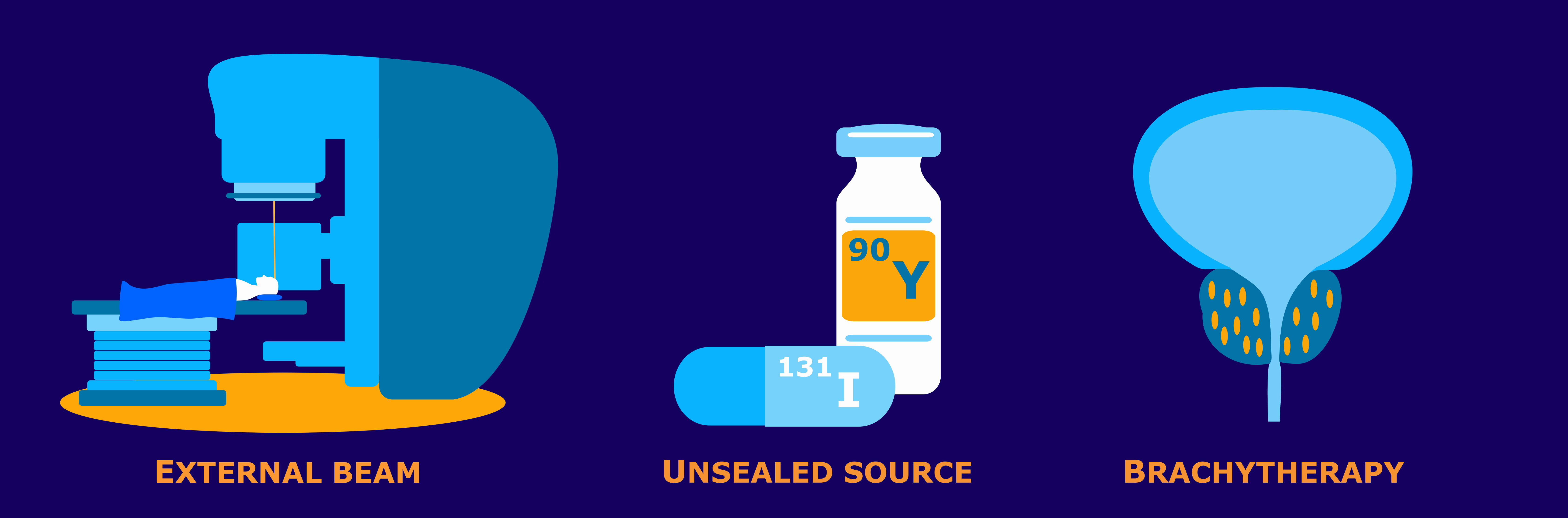
Radioactivity defines the release of ionizing radiation from a substance. Whether it emits alpha or beta particles, gamma rays, x-rays, or neutrons, the radioactivity of a material is a measure of how many atoms within it decay over a specific period of time. The curie (Ci) and becquerel (Bq) units quantify radioactivity.1


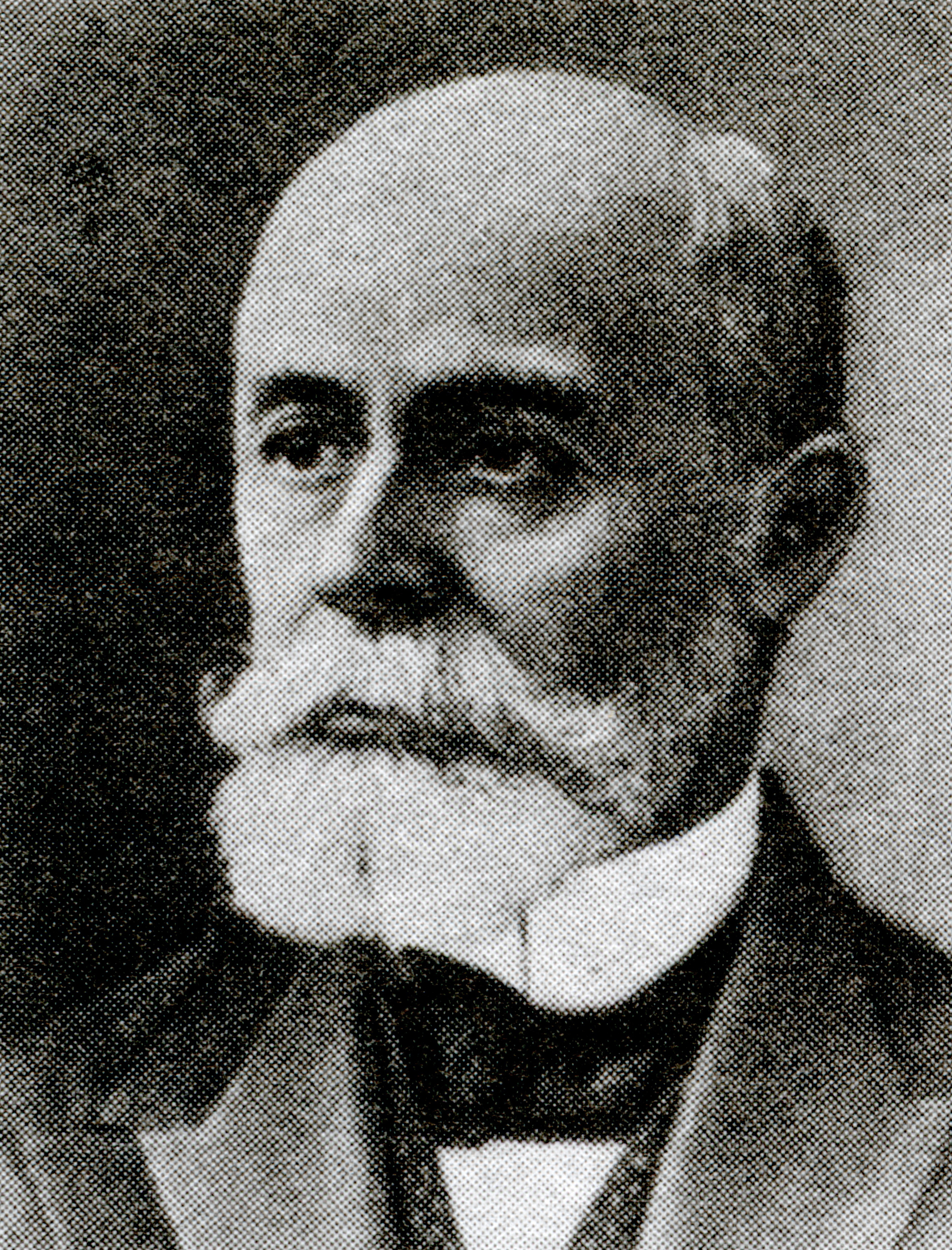
Antoine Henri Becquerel was a French physicist, engineer, and Nobel laureate who discovered evidence of radioactivity. Becquerel’s earliest works centered on the subject of his doctoral thesis: the plane polarization of light, with the phenomenon of phosphorescence and absorption of light by crystals. Early in his career, Becquerel also studied the Earth’s magnetic fields. In 1896, Becquerel discovered evidence of radioactivity while investigating phosphorescent materials such as some uranium salts. For his work in this field, he shared the 1903 Nobel Prize in Physics with Marie Curie and Pierre Curie. The SI unit for radioactivity, becquerel (Bq), is named after him.2

The curie (Ci) unit was created in 1910 by the International Congress of Radiology to measure radioactivity. Pierre Curie, another French physicist, and his wife Marie Curie, who also sat on the committee that named the unit, were the inspirations for the name through their radioactive studies. The original definition of the curie was “the quantity or mass of radium emanation in equilibrium with one gram of radium (element)”.3 In 1975, the becquerel replaced the curie as the official radiation unit in the International System of Units (SI) where 1 Bq = 1 nuclear decay/second4. The relationship between the two units is 1 Ci = 37 GBq (giga becquerels).

Exposure quantifies the extent of radiation going through the atmosphere that reaches a person’s body or a material. Numerous radiation monitors gauge exposure, utilizing the units of roentgen (R) or coulomb/kilogram (C/kg).1

The roentgen is a legacy unit of measurement for the exposure of X-rays and gamma rays. This unit is defined as the electric charge freed by such radiation in a specified volume of air divided by the mass of that air and has the value 2.58 x 10⁻⁴ C/(kg air).5 It was named after Wilhelm Roentgen, a German physicist who discovered X-rays and was awarded the first Nobel Prize in Physics for the discovery.

In 1928, the roentgen became the first international measurement quantity for ionizing radiation defined for radiation protection. This is because it was, at the time, the most easily replicated method of measuring air ionization by using ion chambers.6 However, although this was a major step forward in standardizing radiation measurement, the roentgen had a disadvantage: it was only a measure of air ionization rather than a direct measure of radiation absorption in other materials, such as different forms of human tissue. As a result, it did not take into account the type of radiation or the biological effects of the different types of radiation on biological tissue. Consequently, new radiometric units for radiation protection came to be which took these concerns into account.7

The SI unit for measuring exposure to ionizing radiation is coulomb per kilogram (C/kg). Interestingly, unlike other SI radiation units, this unit does not have a specific name. It officially replaced the previous unit, the roentgen, in 1975, with a transition period of at least ten years.8 The SI unit of electric charge, the coulomb, was named in honor of Charles-Augustin de Coulomb in 1880. Charles-Augustin de Coulomb was a French physicist whose best-known work is his formulation of Coulomb’s law. This law states that the force between two electrical charges is proportional to the product of the charges and inversely proportional to the square of the distance between them. He also made important contributions to the fields of electricity, magnetism, applied mechanics, friction studies, and torsion.9

Absorbed dose refers to the quantity of energy that is absorbed by an object or person where the energy is deposited by ionizing radiation as it passes through materials or the body. The units, radiation absorbed dose (rad) and gray (Gy), measure absorbed dose.1

In 1953, the International Commission on Radiation Units and Measurements (ICRU) adopted the unit rad at the Seventh International Congress of Radiology. This was the unit that replaced the rep, roentgen equivalent physical (detailed later in this blog). Although many believe that the rad is an abbreviation of “radiation absorbed dose”, the ICRU never identified it as such. This suggests that the term “rad” was as a standalone word to be a unit for absorbed dose. There was no documented discussion regarding the use of the rad prior to the Seventh International Congress of Radiology. The closest discussion was during the meeting in 1951 when they determined the need for this type of unit. In 1975, the gray (Gy) replaced the rad as the SI unit of absorbed dose where 1 Gy = 100 rad.10

Louis Harold Gray was a 20th century English physicist who worked mainly on the effects of radiation on biological systems. He was one of the earliest contributors to the field of radiobiology. He worked as a hospital physicist at Mount Vernon Hospital in London and developed the Bragg–Gray equation in collaboration with the father and son team of William Henry Bragg and William Lawrence Bragg. Bragg-Gray theory is the basis for the cavity ionization method of measuring energy absorption by materials exposed to ionizing radiation. Gray’s contributions to radiobiology were numerous. Amongst many other achievements, he developed the concept of RBE (Relative Biological Effectiveness) of doses of neutrons and initiated research into cells in hypoxic tumors and hyperbaric oxygen.11 Gray defined a unit of radiation dosage (absorbed dose) which was later named after him as an SI unit, the gray.

Dose equivalent, also known as effective dose, is a measurement that combines the amount of radiation absorbed and the impact it has on the human body. When it comes to beta and gamma radiation, the dose equivalent is equal to the absorbed dose. However, for alpha and neutron radiation, the dose equivalent surpasses the absorbed dose because these types of radiation have a greater biological impact resulting from their increased ability to damage tissue. To quantify dose equivalent, we use the units of roentgen equivalent man (rem) and sievert (Sv).1

Roentgen equivalent man, or “rem”, was first proposed for use in 1945, but under a different abbreviation. The roentgen was the only unit capable of expressing a radiation exposure at the time. However, it fell short being specifically measurable for photons. Workers came into contact with many other forms of radiation such as alpha particles, beta particles, and neutrons, so Herbert Parker, British-American physicist, created units that would be able to gauge exposures to many types of radiation. These were the roentgen equivalent physical (rep) and the roentgen equivalent biological (reb). Due to similarity in pronunciation between rep and reb, reb was eventually renamed to roentgen equivalent man or mammal (rem) to avoid confusion.

The first appearances of the rem unit in scientific literature were not until 1950.10 The rem related to the rad by multiplying the latter by a quality factor (QF) used to account for the varying biological effects of the different types of radiation. The rad in turn may be obtained from the roentgen by multiplying a dose conversion factor. In air, the dose conversion factor relationship between rad and roentgen is 1 R = 0.88 rad. The absorbed dose to a material is in turn found by multiplying 0.88 by the ratio of the mass energy absorption of the material to that of air.12

Rolf Maximilian Sievert was a Swedish medical physicist. He is best known for his work on the biological effects of ionizing radiation and his pioneering role in the measurement of doses of radiation, especially in its use in the diagnosis and treatment of cancer.13 Sievert contributed significantly to medical physics, earning him the title of “Father of Radiation Protection”. The sievert (Sv), the SI unit representing the stochastic health risk of ionizing radiation, is named after him. The sievert officially replaced the rem as the international SI unit in 1979 with 1 Sv = 100 rem.14

Conclusion

In summary, the history of radiation units is a journey that reflects the progress in our understanding of radiation’s properties and its impact on living organisms. The development of these units has enabled safer and more accurate measurement and assessment of radiation exposure and its effects on human health. By understanding the intricate relationship between radiation and our bodies, we can now take proactive measures to mitigate its harmful effects and promote a safer environment for all.

Sources

  1. NRC: Measuring Radiation. Nrc.gov. Published 2017. https://www.nrc.gov/about-nrc/radiation/health-effects/measuring-radiation.html
  2. The Nobel Prize. The Nobel Prize in Physics 1903. NobelPrize.org. Published 2019. https://www.nobelprize.org/prizes/physics/1903/becquerel/biographical/
  3. Curie – Unit of Radioactivity | nuclear-power.com. Nuclear Power. https://www.nuclear-power.com/nuclear-engineering/radiation-protection/units-of-radioactivity/curie-unit-of-radioactivity/
  4. ‌ Bell DJ. Becquerel (SI unit) | Radiology Reference Article | Radiopaedia.org. Radiopaedia. https://radiopaedia.org/articles/becquerel-si-unit
  5. ‌ Bashir U. Roentgen (unit) | Radiology Reference Article | Radiopaedia.org. Radiopaedia. Accessed August 29, 2023. https://radiopaedia.org/articles/roentgen-unit?lang=us
  6. ‌ Roentgen – Unit of Exposure | nuclear-power.com. Nuclear Power. Accessed August 29, 2023. https://www.nuclear-power.com/nuclear-engineering/radiation-protection/radiation-exposure/roentgen-unit-of-exposure/
  7. ‌ Roentgen (unit) explained. everything.explained.today. Accessed August 29, 2023. http://everything.explained.today/Roentgen_(unit)/
  8. ‌ Bell DJ. Coulomb per kilogram | Radiology Reference Article | Radiopaedia.org. Radiopaedia. Accessed August 29, 2023. https://radiopaedia.org/articles/coulomb-per-kilogram
  9. ‌ Laboratory NHMF. Charles-Augustin de Coulomb – Magnet Academy. nationalmaglab.org. https://nationalmaglab.org/magnet-academy/history-of-electricity-magnetism/pioneers/charles-augustin-de-coulomb/
  10. ‌ Why Did They Call It That? The Origin of Selected Radiological and Nuclear Terms. Museum of Radiation and Radioactivity. Accessed August 29, 2023. https://orau.org/health-physics-museum/articles/selected-radiological-nuclear-terms.html#rad
  11. ‌ LH Gray Memorial Trust: About L.H. Gray. www.lhgraytrust.org. Accessed August 29, 2023. http://www.lhgraytrust.org/lhgraybiography.html
  12. ‌Dosimetric Quantities and Units. U.S. NRC. Published October 25, 2010. Accessed September 11, 2023. https://www.nrc.gov/docs/ML1122/ML11229A688.pdf
  13. Aip.org. Published 2023. Accessed August 29, 2023. https://pubs.aip.org/physicstoday/Online/8433/Rolf-Sievert
  14. ‌ Bell DJ. Sievert (SI unit) | Radiology Reference Article | Radiopaedia.org. Radiopaedia. https://radiopaedia.org/articles/sievert-si-unit?lang=us
30 Jun 2023
Diagnostic and Radiation Therapy Examples

Other Forms of Radiation Therapy and Diagnostic Machines

In our last blog post, we explored the timeline of diagnostic imaging and radiotherapy developed using x-rays. It was the original discovery of x-rays by Röntgen that led to the creation of these medical practices. However, other researchers of the 20th century also invented diagnostic tools and radiation therapy without x-ray involvement at all. We will explore a few of these imaging and radiotherapy machines or procedures in this blog.

Diagnostic Imaging

MRI

One of the most well-known imaging procedures is MRI, which stands for Magnetic Resonance Imaging. Research for MRI began early in the 1970s, but investigation of the magnetic resonance principles started as early as 1945. By happenstance, Felix Bloch, a Swiss physicist working at Stanford, and American physicist, Edward Mills Purcell at Harvard, conducted—nearly simultaneously—an experiment for a new mode of nuclear induction. This led to published papers from both physicists on nuclear magnetic resonance. After publication, both Purcell and Bloch won the 1952 Nobel Prize for this research.1

The process to produce 2D images in an MRI using nuclear magnetic resonance (NMR) was discovered by Paul Lauterbur and Peter Mansfield. Lauterbur published the first nuclear magnetic image in 1973, eventually producing 3D images as well. However, he still had long to go before these images would be practical for application with actual patients. Mansfield continued to expand on Lauterbur’s work, developing the echo-planar imaging technique to improve image quality and scan time in the late 1970s.  By 1977, physician Raymond Damadian created the first full body MRI machine. Through continuous improvements and further research, today’s MRI provides a safe, effective way of capturing images in the brain, portions of the body, the cardiovascular system, and central nervous system. MRIs provide information on size, location, classification, and grade of lesions so as to aid diagnosing stroke, MS, blood supply issues, and any brain damage.2

Ultrasound

Today, ultrasound is most widely known in the OB/GYN field of medicine. Through its invention, however, this diagnostic tool proved its range of utility before becoming a staple in wellness checks during pregnancies. The origin of ultrasonography is often credited to Lazzaro Spallananzi, a physiologist who discovered echolocation throughout 1794 from various experiments involving bats. The principles that serve as a basis for echolocation are also those functioning for medical ultrasound technology today. In 1942, neurologist Karl Dussik first used ultrasonic waves in a diagnostic procedure while attempting to find brain tumors. He later published a report of musculoskeletal ultrasonography in 1958, laying the groundwork for diagnostic musculoskeletal ultrasound.3

Professor Ian Donald from the University of Glasgow was responsible for the development of medical ultrasound in clinical practice. During his research from the late 1950s into the 1960s, there was clinical skepticism about the use of ultrasound. Many doctors felt that manual abdominal and pelvic examinations had proven adequate for medical diagnoses. With his co-workers, Donald performed several studies to show the likelihood of misdiagnoses of a cyst versus a malignant mass. By publishing the findings in 1958, this became a critical point in time for encouraging use of the medical ultrasound. Donald and his colleagues eventually developed an automatic scanner that utilized a full bladder for early pregnancy detection in 1963. This set the path to the now common practice of ultrasounds in the OB/GYN field today.4

PET Scans

Experimental physicist, C.D. Anderson, observed particles from cosmic rays in 1932 that were the same mass as an electron, but which moved in a strong magnetic field along a path opposite to that of an electron indicating a positive charge. He named these particles as “positrons” or positive electrons.

Emission of these positrons was the proof that Irène Joliot-Curie and Frédéric Joliot-Curie used to prove the artificial creation of radioactive elements to win the 1935 Nobel Prize in Chemistry. The creation of these artificial elements was the main focus over the following years due to their significance to military research from the late 1930s until the 1950s. Imaging studies began taking strides as a result of the desire by Louis Sokoloff, an American neuroscientist who hoped to relate mental function to brain function. This encouraged research through the 1950s until the first tomography unit attempt in 1968. This was the turning point in which PET scan development accelerated into the machines we are more familiar with today.5

PET scans can provide imaging through the detection of the gamma ray pairs that are created when a positron annihilates with an electron inside the tissue where the positron emitting isotope has collected. Whereas other diagnostic scans capture the form of an object, PET scans correlate with the metabolic functions of the tissues that uptake the radioactive compound. The compound with the radioactive tracer is typically a form of glucose called FDG (fluorodeoxyglucose), which is used as an energy source by cells and is given to the patient by means of an IV injection. The more rapid the metabolism of the cells in a tissue, as you have with cancer cells, the more uptake of the FDG. This differential uptake is then seen as an enhanced region on a PET scan. 5

Radiation Therapy

Gamma Rays – Gamma Knife (and Cobalt Therapy)

Gamma radiation from radioactive cobalt sources has been harnessed to create an alternative method for producing therapeutic radiation beams. Similar in design to the previously discussed LINAC machines, a Cobalt unit uses a single Co-60 source to produce the radiation beam for treatment. A machine of a fundamentally different design, the Gamma Knife was developed for the treatment of brain tumors. The Gamma Knife is a stereotactic radiosurgery technique that allows for functional neurological surgery. This machine uses anywhere from 192 to 201 C0-60 arrayed in a semi-spherical shield with each source collimated to point their radiation to the same point in space. This type of precision allows for the potential to treat brain tumors and, in turn, pain, movement disorders, and even some behavioral disorders for patients who had not been responsive to other treatment.6

The Gamma Knife was created through the inspirations of Swedish professors Borje Larsson and Larks Leksell in the 1950s. After an investigation using proton beams with stereotactic devices capable of pinpointing targets in the brain, the researchers eventually gave up on that approach due to cost and complexity. Motivated to find an alternative, Larsson and Leksell went on to create a prototype Gamma Knife system in 1967. The prototype unit was a success, used in Sweden for twelve years  and led to a second Gamma Knife in 1975, before other units began appearing globally in the 1980s.6

Proton Therapy

Protons were discovered in the early 1900s. New Zealand physicist Ernest Rutherford’s research during this time led to his discovery of a nuclear reaction that led to the “splitting” of an atom, where he found protons. As a charged particle, the protons have a finite range in matter. The proton’s interaction with matter produces ionization as the proton slows down along its path, losing energy. A peak of deposited dose then occurs at a depth that is proportional to the proton’s original energy.7

The cyclotron, invented by the American physicist Ernest O. Lawrence along with his associates in 1929, proved an efficient way to produce beams of protons. The cyclotron was capable of accelerating protons to high enough energy that, towards the middle of the 20th century, it was suitable for application in cancer treatments.

Dr. Robert Wilson, another American physicist, was the one to spark the idea of proton therapy for battling cancer. He wrote a seminal paper on the functionality of particle beams (comprised of either protons or other heavy-charged particles) and their ability to disperse their energy in the body—the initial release would be small, then exponentially grow when the beam reaches the end of its path for maximum effect against the tumor. Dr. Wilson published the paper in 1946 and received credit to being the inspiration of further research on proton therapy and the continued development of cyclotrons.7

Brachytherapy and Interstitial Therapy

Brachytherapy is an internal radiation therapy technique involving direct placement of radiation inside a patient’s body cavity. This procedure involves small, radioactive source implants that are generally positioned in a body cavity to be near a cancerous tumor. The radioactive material, typically encapsulated within suitable housing material such as titanium, constantly exposes the tumor to a stream of radiation until removal of the radioactivity. These implants will be temporary, and the patient is either hospitalized or kept in a special suite for the duration of the treatment.

Another form of therapy, interstitial therapy (or interstitial brachytherapy), uses sources that are placed directly into the tissue. These may either be temporary or permanent implants of small, encapsulated sources on the order of the size of a rice grain. For treatments with permanent implants, it is safe enough for the patient to go home with a few simple safety guidelines to follow.8

Brachytherapy was first used in 1901 during an attempt to treat lupus. Alexandre Danlos and Paul Bloch completed this treatment with a radioactive sample from Marie Curie. Shortly after, in 1903, Margareth Cleaves used brachytherapy to treat cervical cancer. It became a popular radiotherapy technique to treat breast, cervical, and prostate cancer. As technology advanced along with the use of brachytherapy through the 1900s, imaging-guidance became a valuable asset. More precise dosimetry became possible and allowed for better brachytherapy planning when doctors could use diagnostic modalities such as CTs, MRIs, or ultrasounds. Today, brachytherapy is a more popular treatment for conditions ranging through gynecological, genitourinary, ocular, and head & neck cancers.9

Summary and Conclusion

The 20th century saw tremendous growth and development in diagnostic machinery and radiotherapy. From soundwave technology that gave way to ultrasound, magnetic resonance inspiring MRIs, and the discovery of positrons, medical and non-medical fields can run diagnostic imaging that suits their needs exactly for the most effective information. Radiation therapy research continuously improves the technology and practices used for the wellbeing of patients. Precision instruments such as the Gamma Knife, Cyberknife, and modern LINACs permit for types of treatments that would otherwise be impossible whilst at the same time reducing the side effects associated with external beam therapy. Proton therapy calculations allow for precise travel through the body for maximum effect against cancerous tumors. Brachytherapy provides a treatment option that directly targets tumors without having radiation travel through the body to the target tissue. Modern image guidance ensures source placement exactly where treatment is necessary and that externally directed therapy beams hit their target. As these modalities evolve, we witness the growing reality of safer, more effective diagnoses and cancer treatments within our lifetimes.

The Versant Physics team has experience that covers a range of equipment. This includes dental units, mobile c-arms and Cone-beam CTs, as well as high energy LINACs and even Proton Therapy units and Cyclotrons. To learn more about how our services can help you, contact us to set up a meeting.

Sources

1. NMR Basics | Nuclear Magnetic Resonance Spectroscopy Facility | University of Colorado Boulder. www.colorado.edu. https://www.colorado.edu/lab/nmr/nmr-basics

2. The History of the MRI – DirectMed Parts & Service. https://directmedparts.com/history-of-the-mri/

3. D. Kane and others, A brief history of musculoskeletal ultrasound: ‘From bats and ships to babies and hips’, Rheumatology, Volume 43, Issue 7, July 2004, Pages 931–933, https://doi.org/10.1093/rheumatology/keh004

4. History of Ultrasound – Overview of Sonography History and Discovery. Ultrasoundschoolsinfo.com. Published December 27, 2021. https://www.ultrasoundschoolsinfo.com/history/

5. Henry N. Wagner, A brief history of positron emission tomography (PET), Seminars in Nuclear Medicine, Volume 28, Issue 3, 1998, Pages 213-220, ISSN 0001-2998, https://doi.org/10.1016/S0001-2998(98)80027-

6. History and Technical Overview | Neurosurgery. Neurosurgery. Published 2018. https://med.virginia.edu/neurosurgery/services/gamma-knife/for-physicians/history-and-technical-overview/

7. History of Proton Therapy – NAPT. NAPT. Published 2018. https://www.proton-therapy.org/about/history-of-proton-therapy/

8. Radiation Oncology. Radiation Answers. Accessed May 31, 2023. https://www.radiationanswers.org/radiation-sources-uses/medical-uses/radiation-oncology.html

9. Mayer C, Kumar A. Brachytherapy. PubMed. Published 2021. https://www.ncbi.nlm.nih.gov/books/NBK562190/

21 Apr 2023

X-Ray Evolution: An Accident that Revolutionized Healthcare

It is no question that modern diagnostic imaging and radiation therapy machines play large parts in today’s healthcare. But where did x-ray usage begin? Let’s journey through the timeline of when the first x-ray was taken to today and the growth of its technology into some of the most common machines you may see for diagnostic imaging and radiation therapy.

Diagnostic Imaging

1895

Wilhelm Conrad Röntgen, a physicist, became the first person to observe x-rays, and quite accidentally. Röntgen noticed a glow coming from a nearby chemically coated screen while testing whether cathode rays (known today as electron beams) could pass through glass. After examining the rays emanating from the electrons impacting the glass and their unknown nature, Röntgen dubbed them as “X-rays”. He learned that x-rays could penetrate human flesh but less so higher density substances like bone or lead, and that they were photographable.1

Röntgen’s work paved the way for x-rays to become a vital diagnostic tool in medicine. Dentists such as Otto Walkhoff, Frank Harrison, and Walter König helped spark the advancement of dental radiography, publishing their findings on the topic in 1896.2 During the Balkan War in 1897, doctors used the rapidly improving x-ray radiographs to find bullets and broken bones inside patients on the battlefield.1

1900-1950

Fluoroscopy was invented after the discovery of x-rays in 1900. Continuing into the 20th century, x-rays and fluoroscopy became popular on a consumer level without the risk fully understood. There were rising reports of burns or other skin damage after x-ray radiation exposure; Thomas Edison’s assistant, Clarence Dally, even passed away from skin cancer in 1904 after working extensively with x-rays. Regardless, products like shoe-fitting fluoroscopes were popular from the 1930s until the 1950s so customers at shoe stores could see the bones in their feet.1

On a medical level, scientists progressed with inventing the modern x-ray tube, introducing film into radiology, and the first mammography. Reseachers performed further studies to get a better understanding of the genetic effects of x-ray exposure and the damage caused. Using fluoroscopes in shops tapered off starting in the 1950’s, the practice proven more dangerous than beneficial.3

1970s

X-rays began transitioning into digital imaging by the 1970s which helped save time, money, and storage space.4 Godfrey Hounsfield of EMI Laboratories created the first commercially available CT scanner in 1972. He co-invented the technology with physicist Dr. Allan Cormack and both researchers were later on jointly awarded the 1979 Nobel Prize in Physiology and Medicine.

The cross-sectional imaging, or “slices”, from CT scans made diagnosing health issues like heart disease, tumors, internal bleeding, and fractures simpler for doctors while also being easier on the patients. Through the following years, with how effective the CT scanners proved to be improvements on the design were quickly developed.5

1990s

In the early 1990s, developments with x-ray computed tomography came into a new realm of imaging. Newer CT scanners allowed for the X-ray source to scan in a continuous spiral around the body, which gave an image of a whole organ at once instead of individual cross sections.5 This technique is called helical or spiral CT scanning. C-arm machines created by companies such as Philips introduced Rotational Angiography, in which the C-arm takes a series of images around the patient. This generates an almost 3D picture when viewing the images in a loop.6

Diagnostic Imaging of Today

Diagnostic imaging from the 2000s to present day demonstrate enhanced versions of all diagnostic tools from history. Doctors still use general x-rays for determining severity of injury, check disease, and to evaluate treatment. CT scans have continued to develop so that analysis of issues occurring in a patient’s body can be more clearly seen to determine treatment.7 Mammograms have become instrumental in determining early diagnosis of breast cancer; fluoroscopy still helps doctors today determine effect of movement to certain areas of the body–although not to determine if you should buy a pair of shoes.

Within dental care, veterinary work, and even unexpected industries such as manufacturing and mining, diagnostic imaging has proven its worth.

Radiation Therapy

1895 – 1910

The discovery of x-rays also ushered in research on radiation treatment, eventually leading to some of the machines we see in today’s healthcare. Use of x-ray treatments began between 1895 and 1900, generally delivered as singular, large exposures to the necessary areas. Despite the high doses, the treatments proved to be insufficient over the area of exposure for actual treatment of malignancies, appearing to cause extensive damage to normal surrounding tissues instead.8

1911

As damage from radiation to healthy tissues became more widely known as an issue within the medical world, solutions to a more controlled type of treatment came to creation in 1911. Around this time external beam radiotherapy (XRT) and slow, continuous low-dose-rate (LDR) radium treatments were established from the studied principle of fractionation.8 Splitting the total radiation dose into smaller fractions and delivering it over days achieved better management over cancer growth even while reducing adverse effects to non-cancerous areas of the body.

1920 – 1930

The Coolidge tube, initially invented by William D. Coolidge in 1913 as an improvement over the earlier Crookes tube methods to produce x-rays, became the most popular method for x-ray generation during the 1920s. The Crookes tube used a high voltage between an anode and a cathode, depending on the ionization of the gas in the tube for x-ray production. The Coolidge tube introduced a hot cathode that directly produced electrons from its surface allowing for better control of both energy and intensity of the x-rays produced. Coolidge tubes produced x-rays usable for both diagnostic and therapeutic purposes.

1930 – 1950

Development of more refined machinery truly began within the 1930s for radiation therapy. One of the first machines used for superficial therapy was the Grenz Ray machine. Dr. Gustav Bucky (inventor of the first x-ray grid as well in 1913), invented and made this machine in the 1930s for commercial use by German company Siemens Reiniger. The Grenz Ray machine was effective as a treatment for skin cancers (superficial cancers) due to the rays not passing deep into the body.9 Bucky labeled the rays Granz, the German word for border, as their biological effects seemed to be bordering those of UV light and traditional x-rays.

Treatments for delving deeper started seeing further progression by 1937 through development of supervoltage x-ray treatment; this dubbed the time period from 1930 – 1950 as the “Orthovoltage Era”. Supervoltage x-ray tubes had a staggeringly high kilovolt range compared to previous machines (50 kV to 200 kV). These orthovoltage treatment machines provided higher and variable energies to treat deeper tumors.8 Continued advancements in the Orthovoltage Era became the beginning of electron beam therapy.

1950 – 1980

From the rise in supervoltage x-ray treatment and through inspiration from other machines such as the Grenz Ray machine emerged the linear accelerator. Also known as a LINAC, these machines nowadays are large devices that emit high energy x-rays and electron beams. After use of the first linear accelerators to treat cancer patients in 1953–with lower energy and restricted range of movement–English physicist Frank Farmer developed a comparison between them and other popular machines of the time. This included resonant transformer units, van de Graaff units, and gamma ray units like Co-60 radiation machines.10

In 1962, the conclusions Farmer came to were that LINACs were the ideal choice for larger departments due to a higher price point and hefty size. However, its design at the time left something to be desired. His hopes for linear accelerator improvements included a more robust operation, smaller unit size, and better vacuum systems. Through manufacturers’ dedicated work on improvements up into the 1970s, the LINAC began to fully establish itself as Farmer’s vision came to life with more stable, reliable, compact, and fully isocentric machines.10

Radiation Therapy of Today

After the 1970s, significant changes to linear accelerators slowed. There were no further radical changes to basic structures and concepts for the generation of the x-ray and electron beams. Modern machines became more sophisticated through other improvements. These were advances in beam shaping through the development of multi-leaf collimators, on-board imaging (OBI), and more recently Flattening Filter Free (FFF) designs to permit much greater dose rates. These system and functionality enhancements keep the linear accelerator as the radiation therapy unit of choice for healthcare today.10 Delivering radiation doses deep into cancerous tumors of a patient while minimizing damage to healthy tissue via intensity modulated radiation therapy (IMRT) is achieved confidently with these ever-improving LINACs.

Through the x-ray technologies developed over the last century, scientists continue to develop new methods to safely deliver radiation therapy. Crossing the linear accelerator with the diagnostic imaging capabilities of CT scanners created TomoTherapy®, another radiotherapy option still widely used today. TomoTherapy® machines allow for more precise tumor targeting and preservation of healthy tissue, taking images of cancerous tumors before treatment. The machine will then rotate during treatment to deliver the radiation dose slice by slice, in a spiral-like pattern.11

X-ray radiotherapy is only one type among many cancer treatments now. However, the machines have been honed through the years to create reliable products for healthcare and even veterinary practices. What was once a general, indirect target of radiation exposure has become an extremely pin-pointed, safer technique for the benefit of patients today.

Summary and Conclusion

In summary, x-rays have come a long way since their discovery in 1895 by Wilhelm Conrad Röntgen, revolutionizing medicine. Used for both diagnostic and therapeutic purposes, x-rays underwent significant technological advancements over time starting with producing the first x-ray images on photographic plates. In the early 1900s, the use of x-rays for therapy began with the treatment of skin diseases. By the 1920s, the further developments allowed for the production of higher-energy x-rays. Once in the 1930s, the development of megavoltage x-ray machines allowed for deeper penetration into tissues. Linear accelerators were developed in the 1950s to produce high-energy x-rays and electrons. The 1960s brought the development of computed tomography (CT), which allowed for cross-sectional imaging of the body. In the 1970s, digital radiography was introduced, which allowed for faster image acquisition and processing.

The Versant Physics team has experience that covers a range of equipment including dental units, mobile c-arms and Cone-beam CTs, as well as high energy LINACs and even Proton Therapy units and Cyclotrons. To learn more about how our services can help you, contact us to set up a meeting.

References

  1. History.com Editors. German scientist discovers X-rays. HISTORY. Published July 17, 2019. https://www.history.com/this-day-in-history/german-scientist-discovers-x-rays
  2. ‌Pauwels, Ruben. (2020). HISTORY OF DENTAL RADIOGRAPHY: EVOLUTION OF 2D AND 3D IMAGING MODALITIES.
  3. History Of X-Ray Imaging • How Radiology Works. Published March 12, 2021. https://howradiologyworks.com/history-xray-imaging/
  4. ‌X-Ray Technology: The Past, Present, and Future :: PBMC Health. www.pbmchealth.org. Accessed March 24, 2023. https://www.pbmchealth.org/news-events/blog/x-ray-technology-past-present-and-future#:~:text=Quickly%2C%20military%20doctors%20realized%20the%20life-saving%20potential%20X-rays
  5. Martino S, Cae R, Reid J, Teresa G, Odle B. Computed Tomography in the 21st Century Changing Practice for Medical Imaging and Radiation Therapy Professionals. Published 2008. https://www.asrt.org/docs/default-source/research/whitepapers/asrt_ct_consensus.pdf?sfvrsn=2ac0c819_8
  6. How Philips has been advancing patient care with X-ray for more than a century. Philips. https://www.philips.com/a-w/about/news/archive/standard/news/articles/2020/20201106-how-philips-has-been-advancing-patient-care-with-x-ray-for-more-than-a-century.html
  7. Hanson G. 7 Types of Diagnostic Imaging Tests You May Assist with as a Radiologic Technologist | Rasmussen College. www.rasmussen.edu. https://www.rasmussen.edu/degrees/health-sciences/blog/types-of-diagnostic-imaging/
  8. Cuffari B. The Evolution of Radiotherapy. News-Medical. Published November 11, 2021. Retrieved on March 24, 2023. https://www.news-medical.net/health/The-Evolution-of-Radiotherapy.aspx.
  9. Grenz ray machine used for superficial therapy | Science Museum Group Collection. collection.sciencemuseumgroup.org.uk. https://collection.sciencemuseumgroup.org.uk/objects/co134659/grenz-ray-machine-used-for-superficial-therapy-x-ray-machine
  10. Thwaites DI, Tuohy JB. Back to the future: the history and development of the clinical linear accelerator. Physics in Medicine and Biology. 2006;51(13):R343-R362. doi:https://doi.org/10.1088/0031-9155/51/13/r20
  11. National Cancer Institute. External Beam Radiation. National Cancer Institute. Published May 1, 2018. https://www.cancer.gov/about-cancer/treatment/types/radiation-therapy/external-beam
05 Jan 2023
2023 Happy New Year Banner with blue gradient background

A Year in Review & New Resolutions

Coming full circle to another new year invigorates millions. It is a time to reflect and develop goals for a better self, career, or quality of life. Versant Medical Physics & Radiation Safety also looks eagerly into 2023 and new opportunities of growth. We strive to provide our services to continuously benefit existing or future clients—even while appreciating our building-block actions of 2022. Even as our teams replace calendars in the office and spread poor puns about not seeing each other since last year, we shape our goals to provide exceptional support for healthcare providers to ensure safe workplaces and practices:

Remaining at the Forefront of Medical Physics and Radiation Safety

Sometimes the best resolution is to maintain healthy habits achieved from the year before. Versant Physics will continue its focus on sustaining its status as a trusted, knowledgeable business. Our consulting services demonstrate excellence within medical physics and radiation safety and will continue to in 2023. This involves keeping up with new discoveries in science, seeking value-add opportunities, and ensuring our provided support is top quality. It is with this idea that we strive to keep our competitive edge in all aspects.

Maintaining an edge means aligning ourselves with strong sources when the chances arise. In the past year, Versant acquired Radiological Physics Services, Inc (RPS) and completed a business merger with Grove Physics, Inc. We were excited to welcome Joseph Mahoney from Grove Physics as the new Vice President of Diagnostic Physics. Additionally, Versant brought in the talents of Ray Carlson and his team within RPS. The overall consolidation of these companies’ resources with Versant’s has increased services towards our clients. We are enthusiastic about efficiently using these combined assets to their full potential in 2023.

Another constituent to higher performance levels becoming achievable in the new year is that Versant Medical Physics achieved their ISO/IEC 27001:2013 certification in 2022. This certification demonstrates our dedication to being a trusted source. Not only can we be sought for our expertise in the field, but now to maintain personal information and customer data through even better safeguards in 2023. Being certified for strict security and compliance standards allows for peace of mind to clients using our Odyssey software; the protection of which is performed by our own security management team.

Versant Medical Physics and Radiation Safety ISO/IEC 27001:2013 Certification

As a web-based, modern management system, Odyssey’s enhanced security is not its only feature that is being refined. Odyssey is kept as a radiation software suite that our clients can trust for the central administration of radiation safety programs. This is accomplished by our development team’s dedication to the software’s continuous improvement based off internal and external feedback. Radiation safety programs can quickly become complex and difficult to manage for healthcare companies, large or small. In addition to Versant’s experienced personnel, Odyssey provides clients an all-in-one platform to manage their program more easily and effectively. Within 2023, Versant’s development team will be focusing on projects to publish customizable reports. They will also revamp the centralized audit logging in Odyssey as part of software enhancement requests received through the feedback system.  

Radiation Safety Implementation and Maintenance

Radiation safety has an extensive list of requirements and regulations set through organizations such as the NRC. The necessity of radiation safety programs is unquestionable when working with radioactive substances or ionizing radiation generating equipment. However, the issue remains that implementation and maintenance of these programs can become complicated fast. In 2023, Versant Medical Physics will assist healthcare providers simplify program compliance, protecting their employees and overall business.

Versant provides a variety of services, from dosimetry management to the support of our physicists, Radiation Safety Officers, and specialists. These professionals’ collective years of experience range over key modalities of radiation safety:

  • Any company—regardless of size—can run their badge program through our dosimetry monitoring services. Doing so assures access to our competent technical support team that can accommodate any company’s needs. Dosimetry badge management is top priority for this team to make your program easier to handle. The support team provides technical and customer service to your employees, so they understand best practices for the dosimeters they wear and to simplify compliance. This lets your employees quickly get back to what they do best: providing healthcare to those who need it.
  • Versant Medical Physics has board-certified physicists that cover regulatory and diagnostic services across the board. Versant’s physicists are driven to provide top-tier assistance so that our clients meet regulatory guidelines and ALARA fundamentals easily to protect people: employees, patients, and the general population. We will continue to achieve this in 2023 through provision of full-service support for your company’s radiation safety program’s crucial areas. These services can include but are not limited to equipment testing, radiation shielding and design, and comprehensive audits.

Medical Physics and Radiation Safety Certification and Training Support

Another component of medical physics and radiation safety is requirement (depending on role) of being certified for one’s work. Certifications in this field surround topics such as radioactive material handling in a continually evolving medical field. Our online continuing education training courses are available at any time to earn certifications approved by CAMPEP, AAHP, and ASRT. Many professionals within the medical physics and radiation safety fields need continuing education credits; this can be for compliance purposes or to take on new responsibilities within their company. In addition to providing support for our clients, Versant provides certified courses such as

  • Medical Radiation Safety Officer (MRSO) Training – Compliance knowledge and lectures provided to learn day-to-day requirements for a new Medical RSO. This course has been complimented for its clarity and precision of material.
  • Medical X-Ray Radiation Safety Training – Designed for anyone managing a radiation safety program or working with radiative machines in a medical environment. This course is practical and informative to prepare for any inspection.
  • Fluoroscopy Courses – Safety training that details optimization of fluoroscopy techniques while maintaining ALARA practices. This course has been recognized by previous customers for being comprehensive with employable practices.
  • Department of Transportation (DOT) Training – A combination of safety training for radioactive material transport and general handling. Usable for anyone within the shipping process such as technologists.

Our board-certified physicists are available through online communication to assist with questions or understanding of the content. This ensures that students feel supported through the process. By the end, each student can walk away with an accredited certification for the betterment of their career. Versant Medical Physics will ensure this content reaches as many people as possible to deepen their knowledge base in 2023.

Connecting and Sharing Ideas

Over the last decades, social media became an increasingly significant channel of communication for businesses. As a platform to promote their services and generate brand, companies connect in fashions more popular with the public. Although Versant has seen increases in our reach through social media followings and to the visitors of our website, there are still opportunities to further connect with our fellow companies, clients, and acquaintances within the medical physics and radiation safety fields.

In 2023, Versant Physics will bring a stronger focus into revitalizing our most popular channels for engaging content: our blog and podcast. Versant’s blog is a space for informational posts about radiation in the world and its various practices/safe handling in healthcare, as well as general tutorials on our Odyssey software. With the VersantCast Podcast, hosted by our very own medical physicist, Dr. Eric Ramsay, we take our listeners through various topics surrounding radiation, physics, and healthcare with the expansive knowledge of special guests. We are excited to work back into periodic postings and create subject matter that informs, inspires, and educates both readers and listeners alike.

Versant will also strive to further our network through our most popular social media platforms, being LinkedIn, Facebook, and Twitter. Even as a small company in a niche field, social media gives us the opportunity to connect with other people and businesses within the medical physics and radiation safety industry. Creating spaces to share ideas and new discoveries in science are beneficial to us as well as our followers to further our security in the knowledge surrounding the many fields that handle radiation. To join Versant in our goal to be more connected within the industry, you can follow us on LinkedIn, Facebook, and Twitter.

A Leadership Team that Inspires

Our devoted leadership team’s optimistic goals have shaped the future of Versant Medical Physics since 2016 to bring today’s success. Closing out our list of resolutions, our members of leadership provided what they strive to see to fruition in 2023:

Marcie Ramsay – President, CEO

“As president, I hope to continue providing a positive and supportive workplace environment for our professionals. The new year will also bring the opportunity for me to encourage our team to explore new areas of personal interest and work-life balance through Versant Physics’ recent subscription to the online education platform, MasterClass. On a personal note, I intend to devote more time to daily meditation and reflection.”  

Eric Ramsay – Vice President, Commissioning

“Techniques for treatment in Radiation Therapy get more complex each year. Keeping up one’s knowledge base and gaining expertise in new modalities is challenging with a busy schedule. So, a suitable (and frankly, essential) resolution for the new year will be to focus on continuing education and professional development. This involves staying up to date with the latest research and techniques in the field, attending conferences and workshops, as well as seeking out opportunities for collaboration and networking with other professionals including the staff physicists at Versant. This resolution also includes taking steps to maintain a healthy work-life balance as burn out doesn’t help anyone.”

Ben Ramsay – Vice President, Technology & Finance

“Continue to develop a security mindset. With the increase in cyberattacks globally, and the risks internal and external to Versant, establishing a security-focused mindset is one of our goals in line with our ISO 27001 certification. I will also be focusing on improvement of Odyssey usability for existing clients and ways to bundle the software into our services with non-Odyssey customers that will provide enhanced value. Lastly, Versant will benefit from focuses on cross training staff in 2023 so that we are more flexible and capable of maintaining the highest levels of service possible.”

Joseph Mahoney – Vice President, Diagnostic Physics

“In 2023, I will be aiming for improved frequency and clarity of our client communication. Staying up to date and responsive towards the ever-changing regulatory environment will also allow for a strong start into the new year. Aligning with Versant’s desire for our teams to maintain work-life balances, there will be a strong focus in optimization of physical presence for our staff of physicists in geographic regions only where they are most needed so that they all can get back home more often.”  

Cheers to a productive and exciting 2023!

01 Jun 2022
Patient Wearing Head Coil During MRI Scan In Hospital

Diagnostic Medical Physics in Medicine: Why It’s Important

Many are unfamiliar with the important role that diagnostic medical physics plays in medicine, particularly in the diagnosis and treatment of diseases like cancer.

At Versant Physics, we provide a wide range of diagnostic medical physics services that help healthcare facilities safely and effectively execute procedures for the health and well-being of their patients. Our goal is to help facilities ensure their patients are protected from excessive levels of radiation and that diagnostic equipment is working appropriately, all while maintaining compliance with state and federal regulations.

In this blog post, we’ll break down what diagnostic imaging is, how and why physics principles are applied to diagnostic medicine, and the various roles of a diagnostic medical physicist to help clarify the importance of this profession.

What is Diagnostic Imaging?

Diagnostic Imaging is a range of techniques and equipment used to look inside the body. The purpose of this is to help physicians identify injuries and illnesses, and to help make an accurate diagnosis and treatment plan. This can include a variety of procedures, from simple X-rays for broken bones to more complex procedures involving the brain, heart, or lungs.

Diagnostic imaging procedures are usually painless and noninvasive. However, depending on the test being performed, some patients may be exposed to small amounts of radiation.

diagnostic medical physics

CT scans are a common example of a diagnostic imaging test that emits radiation. In a CT scan, the patient is exposed to a series of X-rays from a variety of angles which are then processed via a computer. The computer creates cross-sectional images of the inside of the body. CT scans are higher-quality images than a normal X-ray and allow physicians to view both hard and soft tissues in the body. They can check for stroke, internal bleeding, chest abnormalities, enlarged lymph nodes, abdominal or pelvic pain, tumors, and more. It is also used to monitor existing diseases such as heart disease and cancer. 

Other common diagnostic imaging procedures include mammography, which helps detect and diagnose breast cancer, fluoroscopy, magnetic resonance imaging (MRI), and ultrasounds.  

Diagnostic Physics and Medicine

Medical physics as a field is divided into five categories, including:

  • nuclear medicine
  • therapeutic medical physics,
  • medical health physics,
  • magnetic resonance imaging physics, and
  • diagnostic imaging.

Diagnostic medical physicists are responsible for ensuring the safe and effective application of radiation used in medical treatments. Specifically, radiology procedures. They work as a member of a patient’s care team, which typically includes physicians, dosimetrists, and radiologic technologists among others.

Equipment Evaluation and Compliance

One of the main roles of a diagnostic medical physicist is to ensure the safe operation of radiation-producing machines and diagnostic radiation detectors. This can include developing imaging equipment specifications, measuring the radiation produced by a piece of equipment prior to clinical use, and proving that the equipment is compliant with regulatory and accreditation requirements.

This also includes assessing all the software, algorithms, data, and computer systems associated with the radiation-producing equipment for accuracy and performance.

Acceptance Testing

Any unit that is used in a diagnostic setting must be periodically reviewed to ensure not only that the image quality is maintained, but that the unit is operating in compliance with the manufacturer’s specifications.

Most states require that a newly installed piece of diagnostic imaging equipment, whether it is brand new or used, be tested by a qualified medical physicist prior to first clinical use. This extremely thorough survey confirms that the unit was installed and set up correctly and ensures that it meets vendor and industry performance standards. It is also an opportunity to identify any potential issues with the unit before it is used on patients.

mammography unit

Typical units that require acceptance testing include fluoroscopic x-rays, radiographic x-rays, PET and PET/CT units, mammography equipment, C-arms, CTs, SPECT cameras, and PACS workstations.

Commissioning

The commissioning process for diagnostic radiation therapy machines such as Linear Accelerators involves testing the unit’s functionality and verifying that dose calculation algorithms work appropriately to produce measured dose calculations.  

Radiation-producing equipment like a LINAC is highly technical and specific. There are many requirements and protocols that detail how this unit should work, from how much energy it produces to the shape and direction of the beam. Diagnostic medical physicists are trained to measure, assess, and implement the optimal baseline values for a unit during the commissioning process.

Patient safety is the end goal of all diagnostic physics commissioning work.

Shielding

Another important aspect of diagnostic physics includes the planning and placement of shielding in areas that use radiation. In the United States, 35+ states require specific shielding designs in any room that houses radiation-producing equipment.

A diagnostic medical physicist can evaluate any shielding that is installed to determine if it will adequately protect workers, patients, and the public from the radiation outside of the scope of a specific treatment. This includes planning for material thickness as well as appropriate placement.

Versant Physics physicists are experienced with a range of equipment shielding requirements, including dental units, Cone-beam CTs, mobile c-arms, high-energy LINACS, Proton Therapy units, and Cyclotrons.

Our team is also experienced with different types of shielding materials, including non-lead materials, which are guaranteed to meet regulatory guidelines and ALARA principles.

Patient Dose & Treatment

Part of a diagnostic physicist’s job is also to ensure the safety of medical imaging modalities being applied in the treatment of individual patients.

They are responsible for determining the exact radiation dose a patient will receive in accordance with the radiation oncologist’s prescription before the patient begins treatment. Creating this therapy plan can take a few hours or multiple days, depending on the complexity of the illness. They also ensure radiation protection guidelines are in place, develop QA tools that ensure optimal image quality, and make sure that all operators are trained in the use of the best imaging techniques.

A diagnostic medical physicist may also monitor the dose of the patient throughout the course of their treatment.

Patients rarely interact directly with the medical physicist on their care team; however, they are a vital part of a safe and effective treatment process.

Versant Physics Diagnostic Support

Our board-certified physicists are able to handle diagnostic physics support for a variety of facilities, including hospitals, clinics, dental offices, and university health systems. With decades of experience, top-of-the-line equipment, and a passion for patient safety, our team is the best choice to assist with your diagnostic medical physics needs.

Contact us for a quote or to learn more about our medical physics support services.

12 Aug 2021

A Step-by-Step Guide to Implementing a Radiation Safety Program

Implementing a radiation safety program is the best way to protect radiation workers and maintain safe radiological conditions in your clinic or university. If you are a new facility starting from scratch, implementing a radiation safety program can be an overwhelming task. We have put together a step-by-step guide to help clarify areas you will need to address.

Who Regulates What?

It is important for any new radiation safety program to understand which regulations to follow. The U.S. Nuclear Regulatory Commission (NRC) is responsible for regulating radioactive materials in the United States. However, they do not regulate radioactive material in any of the 37 Agreement States. These Agreement States have signed agreements with the state’s governor and the chair of the NRC that declare they take responsibility for all radioactive material regulation within the state. Agreement States can set their own rules for how radiation is monitored, handled, and used if they are at least as strict as the NRC.

Each state regulates the use of ionizing radiation generating equipment within the state. It is very important to research your individual state regulations.

For a list of individual state radiation control programs and their specific rules and regulations, we recommend visiting the Conference of Radiation Control Program Directors (CRCPD) website.

Step 1: Identify a Radiation Safety Officer

A Radiation Safety Officer is a required element of a radiation safety program.

According to AAPM Report 160, the RSO in a radiation safety program “is responsible for the implementation, coordination, and day-to-day oversight of the radiation protection program.” An RSO enforces policies and procedures regarding radiation safety and ensures the facility’s use of ionizing radiation is compliant with regulatory requirements, whether that be state or federal. These individuals are required to meet certain education, training, and experience requirements to assume the role.

The responsibilities of the RSO are many. In addition to managing the radiation safety program, this person will:

  • Provide advice and assistance on radiological safety matters,
  • Ensure safe use of radioactive materials,
  • Ensure compliance with regulatory and license requirements,
  • Identify radiation safety problems and correct them,
  • Ensure ALARA practices are enforced,
  • Perform audits and surveys of work areas as necessary,
  • Dose monitoring,
  • Instrument calibration,
  • And more.

Step 2: Get Copies of State and Federal Regulations

Federal regulations can be found on the NRC website. As mentioned above, most states have their own regulatory body. This may also be a good time to contact your state regulator and introduce yourself.

Step 3: Set-up Administrative Documents & QA Program

You will want to lay out the various roles in your radiation safety program in an organization chart. This includes management, IT, radiation safety resources, and additional radiation modalities and departments.

It will also be helpful to create a Standard Operating Procedure Manual on radiation protection that describes emergency procedures, training policies, and credentialing all radiation workers should be familiar with.

Step 4: Establish a Radiation Safety Committee

A radiation safety committee is typically made up of:

  • The RSO,
  • An authorized user of each type of use permitted by the license,
  • A nursing representative, and
  • A representative who is neither an authorized user nor the RSO.

Many universities and larger clinics find an RSC helpful for efficient radiation safety program management. However, they are not always mandatory depending on your use of radiation. You may find a radiation safety committee is not necessary for your facility.

Step 5: X-ray Room Shielding

Radiation Worker Behind Shielding

Facilities that utilize radiation are required to have a shielding plan developed by a qualified expert, such as a medical physicist. Most states also require the shielding plan to be submitted to the state before the equipment can be used.  

When setting up a radiation safety program, it will be necessary to contact an appropriate QE to put together the shielding plan. You will work with them to implement the appropriate materials and signage throughout your facility. Afterward, integrity and regulatory surveys must be performed to ensure compliance with area dose limits.

Step 6: Registration of Radiation Machines & RAM License Application

A new facility with new X-ray equipment must register each unit with the state, typically within 30 days of acquiring the unit. The use of X-ray-producing equipment is regulated on a state-by-state basis. The appropriate forms and required supporting documentation can be found on your state’s regulatory website or by contacting your regulator.

A new facility intending to use radioactive material must apply to either their Agreement State or the NRC for approval. In preparation for submitting the application, all the previous steps should be completed. Many of the items above will be reviewed along with the license application to determine approval status.

Note that some states may require radiation-producing machines to be inspected regularly by state-approved qualified experts to maintain a registration.

Step 7: Set-up a Personnel Monitoring Program

Licensees/Registrants are required to monitor radiation exposure of radiation workers to remain in compliance with occupational dose limits.

Instadose+ Dosimeter

It is important to set up a personnel monitoring program for radiation workers who regularly work with or could encounter radiation while on the job. These programs require personnel to wear a dosimeter badge which measures their total received exposure. RSO’s periodically review the personnel exposures.

There are a variety of dosimeter options available including TLDs, ring badges, and badges that provide on-demand dose reads.

Step 8: Recordkeeping

Implementing a radiation safety program means there will not be existing inspection reports, previous audits, or correspondence with regulators on file to familiarize yourself with. However, as the RSO, you will be responsible for maintaining all records regarding personnel exposure, exposure levels to the public, surveys, calibrations, and any maintenance completed on the facility’s X-ray equipment moving forward. Consult your state regulations to determine how long individual records need to be kept.

Conclusion

While there are many moving parts to setting up a radiation safety program, it is an important aspect of a safe workplace. Following these steps will have you well on your way to leading a successful program.

Our experienced radiation safety officers, health physicists, and medical physicists can help you implement a radiation safety program. Contact sales@versantphysics.com to be connected with a physicist or visit our regulatory page for more information.

Interested in becoming a Radiation Safety Officer yourself? Versant Physics offers a 20-hour online Medical Radiation Safety Officer course that teaches how to implement a successful, compliant radiation safety program. It will help you gain a practical understanding of regulations governing the safe use of radiation-emitting machines and radioactive materials, as well as responsibilities for managing radiation safety in a medical setting.