Author: Versant Physics

13 Oct 2021
Odyssey Inventory Tracking Module

Odyssey “How To” Series: Inventory Tracking Module

Join us for our second interview with Odyssey Implementation Analyst Katelyn Waters, where we discuss how to carry out certain functions of the Inventory Tracking module and answer some of your frequently asked questions.

Odyssey is a radiation safety software suite designed to help RSOs, EHS managers, and Radiation Safety Specialists manage affordable and efficient programs.

https://vimeo.com/622603614

KB 0:09: Welcome to part 2 of our 12-week how-to series highlighting Odyssey radiation safety software. Today we’re back with Odyssey Implementation Analyst Katelyn Waters to talk about the Inventory Tracking module. We’ll be addressing some frequently asked questions we get about the module’s functionality and its use in tracking radioactive materials. Katelyn, can you start by explaining a little bit about the inventory tracking module and its different sections?

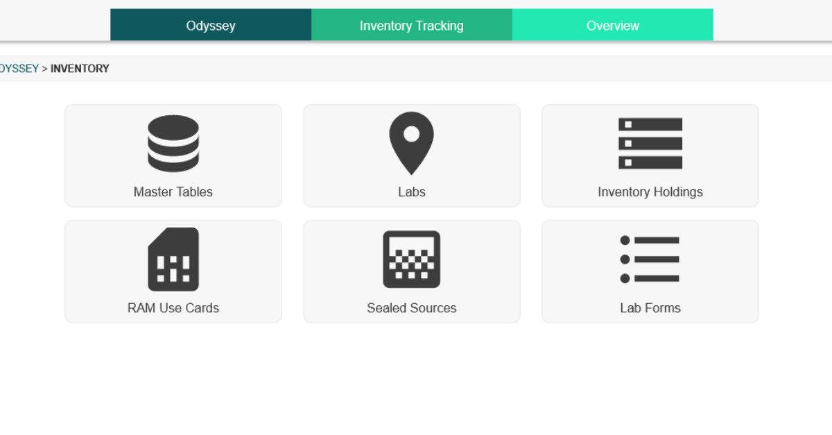
Katelyn 0:35: Absolutely. So let me first go ahead and select the inventory tracking module out of the 12 available modules of Odyssey here. And once I do that, we’re going to have six different sections to look at here. We’re going to have a:

  • Master Tables section which is going to be a master list of any isotopes or compounds you might use in the organization.
  • Labs. We have a section devoted to labs that are going to house radioactive materials. They’re going to each have a profile of information.
  • Inventory holdings are going to be your unsealed sources, each with their own profile as well.
  • We have a RAM Use Card section; this is a really great way to get updates to radioactive material inventories from other users.
  • A Sealed Source section which supports leak testing.
  • And then finally we have a Lab Form section. So this allows you to very quickly fill out forms that relate to any of your labs in this module.

KB 01:30: So, for the section on labs that house radioactive materials, does the area have to be an actual lab?

Katelyn 01:36: That’s a great question. So, we have decided to use the name labs and it really could refer to an actual lab but it may not be as well. It could be an entire building, an entire site, a specific room. So really any location you can designate as a lab.

And if I go in and select this section here we can see the list of available example ones I have for this account. So you can see I have some buildings, I have some overall locations, some warehouses. So, it’s really flexible as to what you can put in as a lab here. And the advantage of doing so is once you add it as a lab it has its own profile, that I mentioned. And I can get to that by selecting the name of any of these here.

So if we go and take a look at Building 1, we have some general information about this building. Like, we can name what this lab is. I can give it a text or number category, a status, I can assign different individuals to it like a health physicist. Importantly, I can update the survey frequency here. And then I have these additional tabs of information. I have a tab that lists out the permits or licenses that relate to this location, maps for it. You can have multiple maps here, say if you want one for each different level of the building or if you want one for a different room.

Survey history, where I can come in and actually select any of these dates if I want more information from the survey for that date, or I can create a new one with this button.

A few other miscellaneous tabs. But the other important one that I want to make sure I point out is this inventory tab. So, this is a big benefit to this section of the module. So any of the locations that you have that house radioactive materials are going to have this tab where it will show you all of the inventory at that location. So you can see I have my unsealed RAM sources here, I have my sealed sources, and then in addition to that, it will also list out equipment and machines at that particular location. It’s really nice to be able to see all of that inventory all in one place.

KB 03:36: That sounds great. So, for the materials that are stored there, does this software do automatic decay calculations?

Katelyn 03:44: Yes. I’ll do an example of one of our sealed sources here. So if I come and take a look at this table we have the name of our sealed sources, a serial number for it, what’s actually contained in terms of the isotope there, the owner or responsible person, if there’s a permit of license that’s authorizing it that’ll be listed out, and then we get into some activity information. And so, when you go and add the sealed source or another radioactive material to Odyssey, to begin with, it’s going to ask you for the known reference date and reference activity. And so that’s what these two columns are. And once that information is in the software, Odyssey is going to use those dates and activities in combination with the half-life of the isotope to always be doing current activity calculations for you on any page refresh. So, all of these are just calculated when I came to the profile here. Like I said, that’s always going to be constantly updating. You can see this information in a few different places, it’s always going to be here on the inventory tab for the different labs, but also in those two separate sections that we already mentioned where you have your unsealed sources and sealed sources too.

KB 04:55: And if I needed to adjust or dispose of any of this material how would I go about doing that?

Katelyn 05:02: Absolutely. You can either go to one of those two sections we talked about, the inventory holdings section or your unsealed, or you can go directly to your sealed sources section. And that will allow you to see a table of different profiles for those materials. I can also just navigate there by selecting anything in these tables. Everything that is possible to be hyperlinked within Odyssey is. So if something you see on the screen has a profile you can usually select that to go to that profile. If I were to choose one of these RAM IDs it’s going to then load that profile of the RAM for me.

This one in particular we have the RAM profile for RAM ID 102 loaded. I can see who the owner or responsible person is, the permits or license that is authorizing it, where it’s at – it’s at building 1 here—what it contains, the compound and isotope, and then all of that activity information that we previously spoke of. Once I’m here, to directly answer your question KB, I have the ability to do some actions on this particular material, and those are found on these buttons in the lower right-hand corner. I can split this RAM, which is going to take this one profile and turn it into more than one, so I can have two, three as many as you want. And you can designate the activity for each of those, so this would be, you know if you’re actually physically splitting this material in real life, and you want that to reflect in your electronic records, you can do it that way.

You can dispose of it, which is one of the actions that you had asked about. So, if you hit this dispose button, it will prompt you to optionally transfer this to waste management. If you do have both inventory tracking and waste management modules, it’s really great for cradle-to-grave tracking there.

You can re-assign this material. That would be to a different owner, a different permit or license, or a different lab. Finally, the other part that you asked about was adjusting the activity. Adjust activity can be done with this button. And like I said, we are automatically calculating these activities, but if you need to manually adjust it for any reason you can do so just by hitting that button.

And anything that you change will show up on this profile as well, that’s what these two fields are where you have an adjusted activity and an adjusted date.

KB 07:17: You mentioned that sealed sources are kept in a separate section for leak testing. How do I view leak test information?

Katelyn 07:24: Yeah, so in order to see that let me actually go to that separate section. I’m going to back out to our main Inventory Tracking section, and this is what we had at the very beginning here of our conversation today with those six different sections. And I’ll go into Sealed Sources where that leak test information is kept. Like I mentioned we do keep these separate due to those leak tests and that workflow, so from this table, I can see which of my sources need to be leak tested. It’s listed in the last column of the table in the Leak Test Required column, and this information comes from when you initially add the source into Odyssey.

If I do choose one, like this one here which has an annual requirement, I can take a look at its profile and we’re going to have some additional information there. On any source that needs to be leak tested, there is a tab called leak test, and this tab allows you to add new leak tests if you want, but it also allows you to view any historical ones that have occurred. So, I can click on this date to get more information if I want, and it will also keep track of when the next one’s due. So this one occurred in May of 2021, and if I come back to my general information tab here, I can see that my next Leak Test is going to be due May of 2022 since this does have an annual requirement. This is really nice that this is automatically going to progress for you once you log a new one on that leak test tab.

KB 08:54: Can I receive reminders for those due dates?

Katelyn 08:56: Absolutely. That’s one of my favorite features of Odyssey is that anything that has a due date associated with a sealed source, machine, equipment, those due dates can push reminders to you which show up as alerts in our system. And they have a post-it note resemblance, they’re going to be in the Alerts and Tasks bar on the lower right-hand corner, and they show up in this format. And you can optionally also get email alerts. Say you’re not logging into this system very frequently; you can have those pushed to your email to make sure you’re definitely aware of those upcoming dates.

KB 09:31: And if I’m being inspected or audited, or maybe I just need to look at records for materials, can I do that?

Katelyn 09:38: Yes. That’s what this History tab is that we have added here that I haven’t spoken about yet. The History tab is going to log any changes that occur for your material, whether that’s going to be an unsealed source or sealed source. You can go back and see these examples here of this Vincent person and what changes he’s made and the date and time those changes were made. So if you need to go back and see when an activity change happened or where a source was, this is a great place to do that for regulatory purposes.

We also have a Documents tab, so if you’ve previously uploaded documents for the source that an inspector might want to see you can always find them here. Say you have a wipe test or some sort of shipment receipt label, you can put that here and always have those to reference.

Finally, say you have an inspector that requests information, but you don’t necessarily want to go through each profile for your sources, we do have a Reporting module which allows you to pull some comprehensive information for everything on the account, and then you can either download that as a CSV file or a PDF and email that to them.

KB 10:44: That all sounds great. And that wraps up our list of frequently asked questions for the Inventory Tracking module. Thanks, Katelyn, for walking through the module with me and clarifying how Radiation Safety Officers can utilize it to manage radioactive material in their radiation safety program. Join us next week for part 3 of our Odyssey How To series where we will be discussing the Reporting module.


Schedule an in-depth demo with our Odyssey team to discuss how the software can assist you with your radiation safety management needs.

06 Oct 2021
Odyssey Personnel Dosimetry module dashboard

Odyssey “How To” Series: Personnel Dosimetry Module

Join us for an interview with our Odyssey Implementation Analyst Katelyn Waters, where we discuss how to carry out certain functions of the Personnel Dosimetry module and answer some of your frequently asked questions.

Odyssey is a radiation safety software suite designed to help RSOs, EHS managers, and Radiation Safety Specialists manage affordable and efficient programs.

KB 00:10: Welcome to Part 1 of our Odyssey how-to series, where we highlight some frequently asked questions about the cloud-based radiation safety software suite and its application in real-world radiation safety programs. Over the next 12 weeks, we’ll be addressing each of Odyssey’s modules, and breaking them down to give a better idea of how they work. My name is KB, and today I’m joined by Odyssey Implementation Analyst Katelyn Waters to discuss the Personnel Dosimetry Module. Thanks for joining me, Katelyn!

Katelyn 00:37: Thanks KB, I’m happy to be here and hopefully we can help answer some of the frequently asked questions that we get for the Personnel Dosimetry module in Odyssey.

KB 00:46: So, the personnel dosimetry module is a key feature of the Odyssey platform. It features customizable widgets and reporting tools that help RSOs and EHS professionals managing a badge program do so in a very efficient way. But how exactly is dosimetry data made available in the module?

Katelyn 01:05: That’s an excellent question, and one of the first that we get whenever we are doing different demonstrations of the software. So, personnel dosimetry does support data from any vendor, but it is primarily designed for Mirion and Landauer. So what it actually does is the software uses a login to either AMP for Mirion or MyLDR for Landauer to view and import that dosimetry information into your account. And so this import happens automatically, we set it up to be daily for Mirion and monthly for Landauer, and so everything’s going to be automatically pulled into the software for you.

KB 01:43: If I have multiple dosimetry accounts can I use this module to manage them?

Katelyn 01:48: Yeah, absolutely. So, it is designed to help aid in the management of multiple accounts. So, whether those accounts are all through one vendor, being Mirion or Landauer, or if they’re from both vendors, this is very beneficial for managing those. You can change the dashboard, that’s what we’re currently looking at here for the module, and this dashboard is a quick at a glance view of your dosimetry account. And you can change what account you’re looking at from this first dropdown menu here that I’m hovering over currently, and you can also change it to look at specific locations or subaccounts, and that’s from the second dropdown menu here.

Additionally, we also have something called a Common Wearer Profile, and what that does is it’s going to combine dosimetry information from more than one account. So if you have a wearer that has a badge from both vendors, or maybe they have two badges that are under different locations or subaccounts, you can combine that information into one profile to make viewing that dosimetry history a lot easier for that wearer.

KB 02:50: So, if I wanted to add or remove any of those multiple badges on my account, can I do so from within Odyssey?

Katelyn 02:57: Yeah, great question. So, the actual addition or editing of any of the badges for wearers for your program will still take place in your vendor portal, whether that’s Mirion, Landauer, or another party. So this particular module is designed to give you some additional features that aren’t available in those vendor portals. Some of those features include ALARA reporting, easy form 5 access, different alerts and reminders, shipping, different management for some of those high-level activities that you’re going to need to do for your program.

KB 03:31: I see. What about this dashboard? Is it possible to change any of the information that’s displayed here?

Katelyn 03:38: Yes. I definitely recommend that when you start utilizing this module in the software that’s something you do right away because it makes it very beneficial for you, and you can do that by selecting this gear icon here, it’s going to take us to show us all of the settings. So each of these boxes we refer to as a widget, and each widget has its own settings for what you want to view on your dashboard. A good example here is the Recently Viewed widget in the top right-hand corner. I have what I want to see available for selection with these checkboxes, I have some drop-down menus for the number of things that I want to see here for the Read activity widget I can choose the time period… so depending on what it is they each have their own settings for these particular widgets, and that way you can really make it for what you need to see for your program.

In addition to that, you can also click and drag things to more prominently display them. So, if I want to have this graph of Read Activity–these are doses that are coming in each day–I can put this at the very top so I can see that a more prominent position. You can also hide things if they aren’t useful to your program. So, say you don’t want this recently viewed widget, I can select the eye icon and hide that and if I were to save these settings it would remove that from my dashboard. So, it’s very customizable.

KB 04:59: You mentioned read activity. Is there a place that I can see who hasn’t read their badge?

Katelyn 05:06: Yeah, so we have a query data section which I can actually go to and show you here. And this section allows you to do some really in-depth searches on your dosimetry data. You can choose to select what you want to search for, so with your example, I would go and search for different personnel, and ones that don’t have a badge reading. And then I come down to this section where it says Include or Exclude and I can choose things that I want to either include in my search or exclude from my search. And for us today, I’ll go ahead and add in an inclusion statement to include people for the current account that we’re looking at, just as an example, and then I can exclude people who have readings and we will only be left with those who do not, so I’ll go ahead and exclude people who have a reading for this year by putting in the date of January 1st.

So we can hit this play button here and it’s going to then search through all of your dosimetry records and give you the corresponding data for your filters. Once it does so, you have a table result at the bottom. And so, the format of this data is a little odd due to the fact that we’re looking at primarily, or not primarily, exclusively demo data. So you can see the employee IDs are very long. But this is what the format of that table would look like for you, just have your own dosimetry data displayed in it.  

And these are linked to other areas of the software. So for these wearers, I can actually select them to go to their profiles, and in addition, we have a list of contact information here that if you were to select this mail icon for it would actually put all of these people into a mailing list for you so you can easily communicate with them. So if we do want to reach out to this group and say hey, you need to please read your badge for 2021, you can do so by this easy feature there.

KB 07:02: Well that seems pretty easy to do. Is there any way for me to receive notification of individuals with high doses?

Katelyn 07:10: Absolutely. So that is actually one of my favorite features that we have added into the Personnel Dosimetry module and it exists in this reporting section. So I’ll go ahead and select that and we’ll take a look at that.

The reporting section has a tab called ALARA. The module has the ability to add in different custom ALARA thresholds. For this particular demo dosimetry account, we have two thresholds added in. I’ve added in an ALARA 1 and an ALARA 2. These are both quarterly thresholds but you can also have them trigger on a single dose, they can be monthly, quarterly, or annually. So, you have a variety of options there. They can also be scoped to particular badge regions that a badge is assigned to. Since these are quarterly thresholds, this next filter of the period gives me the option to choose a quarterly time period but that will change depending on your threshold. And if I do put in last quarter as an example and select run report, what that’s going to do is give me a table of results of individuals that have surpassed that threshold for the chosen time period. So we have these three demo people who have surpassed the ALARA 1 Threshold,  and then one person additionally also surpassed the ALARA 2, so this table is really nice because if they did surpass other thresholds it will let you know that. So if you only want to follow up on the higher of the two you absolutely can do that.

It will tell you the time period that we’re covering currently, and then the cumulative dose for that time period, and then the stage that you’re at in the process. So this will allow you to email each of these individual wearers either notification that they went over this dose threshold or a questionnaire – that form that they actually get sent is completely customizable – and the wearer will receive that via email. Once they do, they will open and view that, if they have any questions they can fill that out, it requires a signature and dates it for them automatically, and then that gets sent back to Odyssey. Once it does get sent back, this pie chart progresses and shows you where this process is. So it will change to being yellow for “waiting for RSO response.” So if you’re an RSO, a radiation safety representative, you can come in here, review that questionnaire, and the responses, and sign off on that to complete the process.

And to get back to your initial question about alerts for these items, throughout this entire process you’re going to be receiving in-software alerts which can optionally be email alerts as well. So when a wearer initially goes over the threshold, any of these thresholds you have set up, you’re going to get an alert that looks like a post-it note like one of these. And then when they fill it out they also will trigger an alert and it’ll let you know they have it filled out so that way you can come in and review that as soon as possible. And so you’re always going to be notified of each stage of the process there as this progresses.

KB 10:07: Well that all sounds great! Thanks, Katelyn, for helping us address some frequently asked questions about Odyssey’s Personnel Dosimetry module. Join both of us next week for part 2 where we’re going to be talking about the inventory tracking module and its ability to assist with tracking radioactive materials in your radiation safety program.

Katelyn 10:26: Thanks, KB.


Schedule an in-depth demo with our Odyssey team to discuss how the software can assist you with your radiation safety management needs, or visit our website to learn more about Odyssey’s radiation safety modules.

01 Oct 2021

How Molecular Imaging and Radiation Therapy Help Fight Breast Cancer

It is estimated that 1 in 8 women in the United States will develop invasive breast cancer within their lifetimes. It is an incredibly devastating disease that affects thousands of people a year. This year alone, 281,550 women in the United States will be diagnosed.

Screening efforts and treatment therapies involving molecular imaging and radiation therapy are key to helping detect and successfully treat breast cancer.

Breast Cancer Statistics

  • Breast cancer is the most diagnosed cancer in women.
  • Breast cancer in women has the highest rate of death compared with any other cancer, besides lung cancer.
  • It is more commonly diagnosed in black women under the age of 45 than white women.
  • Minority women are 72% more likely than white women to be diagnosed with breast cancer before age 50 and are 127% more likely to die of breast cancer before age 50.
  • Men have a 1 in 833 chance of getting breast cancer.
  • In 2021, the World Health Organization reported that breast cancer accounted for 12% of all new, worldwide cancer cases.

Breast Cancer Risk Factors

Age and being born female are the biggest risk factors for breast cancer. Women that have direct relatives with a history of breast or cervical cancers such as mothers, sisters, and grandmothers have a higher risk of developing breast cancer in their lifetimes.

There are several known gene mutations that can be inherited from either parent which grant a higher lifetime risk of developing breast cancer as well.

When functioning correctly, these genes, called Breast Cancer Gene 1 (BRCA1) and Breast Cancer Gene 2 (BRCA2), produce proteins that help repair DNA. These tumor suppressor proteins actually help protect from certain cancers by slowing abnormal cell growth and forcing certain damaged cells to stop working entirely.

However, when present, the BRCA gene mutation can prohibit these proteins from working and building correctly, resulting in cancerous tumors.

By age 80, 55%-72% of women with an inherited BRCA1 mutation and 45%-69% with an inherited BRCA2 mutation will develop breast cancer. People with a BRCA variant also tend to develop breast cancer at a much younger age than those without.

There are also higher instances of BRCA1 and BRCA2 gene mutations in certain racial and ethnic groups. For instance, 2% of Ashkenazi Jewish people carry one of the variants. A study in 2009 determined that black and Latin American women were more likely to have BRCA1 mutations.

Signs & Symptoms

The most common symptoms of breast cancer include a lump or mass in the breast, but physical changes in the appearance of the breast are also reported. This includes skin redness or swelling, bloody or abnormal discharge, thickening of the skin, or scaliness.

Breast cancer can develop without presenting any physical symptoms, however, which is why regular screening and breast exams are so important for prevention.

Screening Recommendations

Regular breast cancer screenings can help discover breast cancer in its early stages before it has spread to other parts of the body, therefore, limiting treatment options and increasing mortality rates. Mortality rates can be reduced by 40% with regular screenings.

Mammography

Mammography is a low-dose x-ray procedure used to detect breast cancer in its early stages, often before a patient has experienced any symptoms like lumps or skin alterations.

This type of x-ray exam exposes the patient to low doses of ionizing radiation to produce an image of breast tissue or the inside of the breast.  

During a mammography procedure, the breast is flattened between two plates on the x-ray unit for several seconds while an x-ray beam is carefully aimed at the area of concern by the radiologist or technologist performing the procedure. It is standard during a normal screening for two views of each breast to be taken. The mammograms are then reviewed by a radiologist, who looks for early signs of cancer or other abnormalities.

Mammograms can also be used if a patient has experienced symptoms of breast cancer and to screen patients who have been previously treated for breast cancer. This diagnostic mammogram includes additional views of the breast not normally taken during a screening.

Radiation received from regular mammograms is cumulative, however, it does not significantly increase breast cancer risk. In the case of screening for cancer, it is more beneficial in the long run to receive a low radiation dose. 

Screening Frequency

Various medical and cancer-related institutions have different guidelines on when it is appropriate or necessary to schedule a mammogram.

The American Cancer Society recommends patients with an average risk of breast cancer between the ages of 45-54 get annual mammograms. Patients aged 55 and older have the option to get a mammogram every other year.

The American College of Radiology (ACR) and the Society of Breast Imaging (SBI) recommend annual mammograms begin at age 40 and continue past age 74.

Molecular Imaging & Breast Cancer

Molecular Imaging is a medical imaging procedure used to help locate breast cancer tumors and determine if cancer has spread to other parts of the body. It is a vital part of the diagnosis and treatment process because it measures biological and chemical processes within the body, compared with regular x-rays which focus on static anatomical images.

Molecular imaging helps physicians determine the appropriate treatment therapies, study the patient’s response to drugs, and closely monitor changes in cellular activity. It is also useful for identifying whether the prescribed therapies are effective and monitoring for reoccurrences.

There are a variety of medical imaging procedures that help visualize chemical processes in the body such as blood flow, oxygen use, or metabolism. Many procedures require an imaging agent such as a radiotracer—a compound containing a small amount of radioactive material—being introduced into the body usually via injection into the bloodstream.

This radiotracer is designed to accumulate in the body in different organs which are then picked up by the imaging device.  It can also attach to different cells or groups of cells and paint a clear picture about precisely where abnormal amounts of metabolic activity are occurring.

PET/PET-CT

Positron Emission Tomography (PET) scans alongside Computer Tomography (CT) are one of the most common molecular imaging technologies used for breast cancer. The combination of these two imaging modalities helps physicians determine the exact location of the tumor, what stage the cancer is at, if it has spread, and what type of treatment will be best moving forward.

In this procedure, a radiotracer that naturally emits positrons as it decays is injected into the bloodstream. These positrons react with electrons in the body and produce energy in the form of photons. These photons are detected by the PET scanner, producing 3D images which show how the radiotracer is being distributed.

On a PET scan, the areas where the radiotracer has accumulated appear brighter and more intense than in the surrounding tissue. This is because cancer cells, when active, absorb more glucose. The higher instance of this metabolic activity is made clear thanks to these “hot spots” on the PET scan.

The PET scan is combined with the CT scan to produce a detailed image of both the patient’s anatomy and the metabolic activity present.

Surgical Treatment Options

In addition to chemotherapy, there are several other treatment options for breast cancer that typically precede radiation therapy.

Lumpectomy

A lumpectomy, also known as a partial mastectomy, re-excision, or biopsy, is a breast-conserving surgery that involves removing part of the breast tissue. The surgery removes the lump or tumor plus a small amount of the healthy tissue that surrounds it.

Mastectomy

A mastectomy is a surgical procedure that removes the entire breast. There are different kinds of mastectomies with varying degrees of severity. The type of mastectomy a patient receives will depend on the stage the cancer is at and if it has spread to the lymph nodes or other areas of the body. 

Radiation Therapy & Breast Cancer

Radiation therapy delivers ionizing radiation particles to specific areas of the body to destroy cancer cells, either as a standalone treatment or in conjunction with other treatment options like surgery. Brachytherapy and External Beam Radiation Therapy are the two most common treatment types.

Brachytherapy

Brachytherapy is a procedure that involves placing small, sealed radioactive material sources inside the body, either directly inside or next to a tumor. Also known as internal beam radiation, this procedure is used to treat cancer by allowing doctors to deliver higher doses of radiation via a needle or catheter to specific areas of the body.

Compared to other types of radiation treatments, brachytherapy is best for cancers that have not metastasized. It is considered as effective as—and sometimes used in conjunction with—external beam therapy. Due to the nature of brachytherapy procedures, there is a smaller chance of radiation exposure to surrounding healthy tissue and organs than with external radiation as it targets the tumor directly.

Because healthy tissue and organs surrounding the tumor are not as affected by the radiation treatment, most people experience few or less serious side effects than occur with external beam therapy. In addition to tenderness, bleeding, or swelling at the treatment area, the side effects a patient could experience depend largely on the type of cancer and therapy being performed. Fatigue is common.

External Beam Radiation

According to the American Cancer Society, external beam radiation is the most common type of radiation therapy used to treat breast cancer. It can be used in both early-stage breast cancer as well as for advanced stages that cannot be removed with surgery.

EBRT normally occurs 3 to 6 weeks after a patient has undergone surgery and/or completed chemotherapy. Small doses of ionizing radiation are delivered to cancer to destroy the cancerous cells. This process is normally a painless outpatient procedure that lasts up to 5 days a week for anywhere from 2 to 9 weeks.

During EBRT, the patient is usually positioned on their back with their ipsilateral arm placed above their head and their shoulder rotated outward. Then, radiation is precisely applied to the area according to the radiation treatment plan.

Throughout the process, a radiation oncologist monitors a patient’s response to the treatment and may alter the prescribed radiation dose or the number of treatments accordingly.

One 2021 study suggests that a lumpectomy plus radiation therapy offers better survival rates than a standalone mastectomy for early-stage breast cancer. Other studies have discovered that the risk of recurrence in a patient who undergoes radiation therapy is between 5% to 10%, while patients who do not receive radiation therapy have a 20% to 40% recurrence rate.

The Radiation Therapy Team

Each patient who undergoes radiation therapy has a dedicated team of radiation professionals on their side who determine exactly how they will be treated. Throughout the treatment process, they also determine if any changes to the radiation treatment plan need to be adjusted.

A radiation oncologist is a specialist in treating cancer with radiation. Their job is to determine which therapy is the best fit for the patient based on their medical history and physical health.

A medical physicist and dosimetrist will also be a part of this team. They work together with the physician to create the treatment plan.

Radiation therapists and technologists are the individuals who physically administer the radiation therapy treatments and operate the equipment. They are the people patients will interact with the most during their treatment.

The Takeaway

There are a variety of available breast cancer treatment therapies including radiation therapy and surgery. Radiation therapies administered in conjunction with chemotherapy and surgical treatment options have a much lower recurrence rate than standalone treatments.

Proper screening and regular exams are the best way to detect breast cancer when it is in its earliest and most treatable stages. If a patient is 40 or older, it is in their best interest to begin scheduling annual mammograms.

In honor of Breast Cancer Awareness month, check out the following organizations providing patient support services and making great strides in research and awareness:

10 Sep 2021
Ore mining

What is Naturally Occurring Radioactive Material?

Naturally Occurring Radioactive Materials (NORM) are just that: materials of natural origin that contain radioactive materials. NORM is found in rock formations, soil, and sand that come out of the Earth’s crust and mantle. This includes elements like radium, uranium, thorium, and potassium, as well as their decay products radium and radon. Many of these elements show up in concentrated areas, like uranium ore bodies, which are then mined for human use.

The Environmental Protection Agency (EPA) defines NORM as “materials that contain any of the primordial radionuclides or radioactive elements… that are undisturbed as a result of human activity.”

Cosmogenic NORM, or cosmic radiation produced by cosmic rays interacting with the Earth’s atmosphere, affects frequent flyers and those who live at higher altitudes.

Learn more about background radiation in our blog post here.

According to the IAEA, the activity concentrations of the radionuclides found in these places are generally low and not considered to be a risk to human health and safety.

Technologically Enhanced Naturally Occurring Radioactive Materials (TENORM)

Human activities such as extraction and processing can expose, disturb, or concentrate NORM. Bringing natural resources from below the ground—in a solid, liquid, gas, or sludge form—and introducing it to the surface brings up the materials that contain radionuclides. When something like this happens, NORM is classified as Technologically Enhanced Naturally Occurring Radioactive Materials or TENORM.

pile of raw coal

It can also concentrate it in some cases. For example, coal-ash, a byproduct of burning coal, contains a higher concentration of NORM than it did when it was mined from the ground. As ash only accounts for about 10% of the weight of unburned coal, the resulting NORM is 10x that of the plant’s coal fuel.

Industries that generate TENORM include:

  • Mining (hard rock/metal, rare earths, uranium, copper, alumina)
  • Energy (oil and gas, coal, fracking)
  • Water treatment (drinking water, wastewater, fish hatcheries)
  • Consumer products (fertilizer, cigarettes, granite countertops, bricks/building materials)
  • Recycling

Region and geology are major factors in the amount of radioactivity and materials such processes can introduce into the environment. When these materials are exposed or concentrated because of industrial processes, humans are exposed to the ionizing radiation they give off. This can result in potential health risks including cancer.

Regulating NORM/TENORM

Radiation levels from NORM are not considered hazardous in the United States.  Therefore, it is not regulated at the federal level.

TENORM is also not regulated by the federal government or the Nuclear Regulatory Commission. It is up to individual states if and how they choose to regulate generation and disposal. Currently, there are 37 Agreement States which regulate NORM within their borders. There is little consistency among different industries and countries regarding NORM. Contact your state’s radiation management branch for more information.

How to Minimize TENORM Exposure

From a radiation safety perspective, some precautions can be taken by professionals and the public to ensure minimal exposure to these radioactive materials. The level of exercised caution ultimately depends on the type of TENORM present. In general, TENORM should be handled only by individuals familiar with radiation safety practices and hazardous industrial substances. Other steps to take include:

  • Implementing a radiation safety program
  • Using appropriate shielding, HEPA filters, and personal protective equipment as necessary
  • Minimizing time spent around TENORM
  • Avoiding eating or drinking around TENORM
  • Minimizing activities like cutting or grinding which can generate dust containing TENORM
  • Properly disposing of TENORM-contaminated waste
24 Aug 2021

The Seven Most Influential Women in Radiation History

The role of women in science is often overlooked. However, the research and discoveries of these brilliant minds have drastically altered commonly held theories in particle physics, chemistry, and nuclear medicine, and contributed to our modern understanding of radiation. In this post, we highlight seven of the most influential women in radiation history and their outstanding accomplishments.

Marie Curie (1867-1934)


Madame Marie Curie was a physicist and chemist whose pioneering research in radioactivity won her two Nobel Prizes in two scientific fields. In addition to her groundbreaking work in nuclear physics and chemistry, she developed the mobile X-ray unit which was first used to diagnose injuries during World War I.

Born in Poland in 1867, Curie moved to France to study physics, chemistry, and math at the University of Paris in 1891. There she met her future husband and research partner Pierre Curie. She earned two degrees from the institution, one in 1893 and another in 1894.

In 1903, Curie and her husband received the Nobel Prize for their joint research in radioactivity alongside Henri Becquerel. They were responsible for the discovery of new elements radium and polonium which came from the radioactive mineral pitchblende, now commonly known as uraninite. She was the first woman to win the Nobel Prize.

In 1910, she was successful in producing radium as a pure metal, further proving the element’s existence, and was awarded her second Nobel Prize in Chemistry in 1911.

Curie served in World War I as the director of the Red Cross Radiology Service. She created small, mobile X-ray units called “Petite Curies” which were vehicles containing an X-ray machine and darkroom equipment. She trained over 150 women to operate the units which ultimately helped treat over one million soldiers near the battlefront.

Curie died in 1934 of aplastic anemia, likely a result of her work with radiation.  

Awards & Recognition

  • 1903 – Received the Nobel Prize in Physics (with her husband Pierre Curie and Henri Becquerel)
  • 1911 – Awarded the Nobel Prize in Chemistry
  • 1920 – Became the first female member of The Royal Danish Academy of Sciences and Letters
  • 1924 – Became an Honorary Member of the Polish Chemical Society
  • Received 4 honorary doctorates from Polish universities
  • The radioactivity unit “curie” is named in honor of Marie and Pierre Curie
  • Element 96 was named curium

Lise Meitner (1878-1968)


Dr. Lise Meitner was an Austrian-Swedish physicist who helped discover the element protactinium-231 and nuclear fission. She received her doctorate in physics—the second woman to do so—at the University of Vienna in 1906. In 1926 she became Germany’s first female professor of physics, a role she held until the rise of Nazi Germany and the Nuremberg Laws forced her to flee to Sweden to escape religious persecution.

She worked closely with Otto Hahn, a prominent chemist, throughout the years. Their work on discovering isotopes resulted in the introduction of protactinium-231.

In 1939, Dr. Meitner coined the term “fission” after discovering that uranium atoms split when bombarded with neutrons. Her role in this major discovery, which allowed for nuclear energy and nuclear bombs, was overlooked by the Nobel Prize committee, and the award was given exclusively to Otto Hahn in 1944. Because of this discovery, she was invited to work on the Manhattan Project, however, she opposed the atomic bomb and declined the offer. She was ultimately nominated for the Nobel Prize 48 times for physics and chemistry projects but never won.

She was a strong supporter of women in science and spent the last half of her life traveling and speaking to female students.

Awards & Recognition

  • 1925 – Awarded the Lieben Prize from the Austrian Academy of Sciences
  • 1944 – Named “Woman of the Year” by the Women’s National Press Club in Washington D.C.
  • 1945 – Became a foreign member of the Royal Swedish Academy of Sciences
  • 1954 – Awarded the inaugural Otto Hahn Prize of the German Chemical Society
  • 1966 – She was awarded the Enrico Fermi Award alongside chemists Otto Hahn and Fritz Strassmann for her “pioneering research in the naturally occurring radioactivities and extensive experimental studies leading to the discovery of fission”
  • 1997- The chemical element meitnerium was named in her honor

Irene Joliot-Curie (1897-1956)


Irene Joliot-Curie was a chemist and physicist known for her work on natural and artificial radioactivity, transmutation of elements, and nuclear physics.  

She was born in Paris, France in 1897 to Marie and Pierre Curie. She studied chemistry at the Radium Institute and completed her Ph.D. in chemistry from the University of Paris. Her doctoral thesis focused on radiation emitted by polonium.

During World War I, Irene worked alongside her mother on the battlefield as a nurse radiographer. For a time, she also taught doctors how to locate shrapnel in soldiers using radiological equipment.

Alongside her husband, chemical engineer Frederic Joliot, Irene studied atomic nuclei. Together they were the first to calculate the accurate mass of the neutron and discovered that radioactive elements can be artificially produced from stable elements. The pair shared the 1935 Nobel Prize in Chemistry in recognition of this discovery, which had practical applications in radiochemistry, specifically in medicine and the treatment of thyroid diseases. In addition, her research on the action of neutrons on heavy elements was an important step in the discovery of nuclear fission.

Outside of her research, Irene was the Chair of Nuclear Physics at the Sorbonne and a Professor in the Faculty of Science in Paris. Beginning in 1946 she served as the director of the Radium Institute and was instrumental in the design of the Institute of Nuclear Physics in Orsay, France. She died in 1956 of leukemia, likely a result of her work with polonium-210.

Awards & Recognition

  • 1935 – Received the Nobel Prize in Chemistry for the discovery of artificial radioactivity (with Frederic Joliot-Curie)
  • 1940 – Received the Barnard Gold Medal for Meritorious Service to Science (with Frederic Joliot-Curie)
  • Was an Officer of the Legion of Honour

Edith Quimby (1891-1982)


Edith Quimby was a pioneer in the field of radiation physics, a founder of nuclear medicine, and is considered the first female medical physicist in the United States.

She was born in 1891 in Rockford, Illinois, and earned degrees in physics and mathematics from Whitman College and the University of California, Berkeley. Much of her early work at the Memorial Hospital for Cancer and Allied Diseases in New York focused on the medical effects of radiation and limiting side effects with proper dosages. Furthermore, she was also interested in the safe application of radioactive isotopes in the treatment of thyroid disease, brain tumors, and other cancers.

Edith Quimby helped found the Radiological Research Laboratory at Columbia University, was the first female physicist president of the American Radium Society and was influential in the founding of the American Association of Physicists in Medicine. She was a professor at both Cornell University Medical College and Columbia University, and she authored several books throughout her career, including the classic Physical Foundations of Radiology (1944), and over 70 scientific papers.

Awards & Recognition

  • 1940 – Recipient of the Janeway Medal from the American Radium Society
  • 1941 – Awarded the Gold Medal of the Radiological Society of North America
  • 1963 – Awarded the Gold Medal from the American College of Radiology
  • AAPM established a lifetime achievement award in her honor

Tikvah Alper (1909-1995)


Tikvah Alper was a renowned radiobiologist and physicist whose work on identifying the infection agent in Scrapie revolutionized scientific understanding of diseases like mad cow disease and kuru.

She was born in 1909 in South Africa and graduated with a distinction in physics from the University of Cape Town in 1929. She was mentored by Lise Meitner as a doctoral student in Berlin from 1930 to 1932 where she published an award-winning paper on delta rays produced by alpha particles.

In addition to her life as a mother and homemaker, she was a physics lecturer at Witwatersrand University and researched in Britain on the irradiation of bacteriophage. She became head of the Biophysics Section in South Africa’s National Physics Laboratory; however, she was forced out of this position in 1951 due to her opposition to apartheid. Afterward, she moved to London with her family and worked her way up to director of Hammersmith Hospital’s MRC Experimental Radiopathology Research Unit in 1962.

Alper found that radiation did not kill the infective agent in Scrapie, an infectious brain disease found in sheep. Instead, by irradiating scrapie samples with different wavelengths of UV light, Alper was able to prove the infective agent was able to replicate despite its lack of nucleic acid. This work became extremely important during Britain’s Mad cow disease outbreak in the 1990s.

Chien-Shiung Wu (1912-1997)


Chien-Shiung Wu, also known as the “First Lady of Physics,” was a Chinese American particle and experimental physicist who worked on the Manhattan project and played an important role in the advancement of nuclear and particle physics.

Madame Wu was born in 1912 in Shanghai. She received a degree in physics from what is now known as Nanjing University and later enrolled at the University of California, Berkeley where she completed her Ph.D. She worked as a physics instructor at Princeton University and Smith College before joining the Manhattan Project in 1944. Her work at the Substitute Alloy Materials Lab was meant to support the gaseous diffusion program for uranium enrichment. Her research also improved Geiger counters for radiation detection.

As a leading physicist on beta decay, Madame Wu was able to confirm Enrico Fermi’s 1933 theory of beta decay. She was also responsible for disproving “the law of conservation of parity” in what is known as the Wu Experiment. In this experiment, she measured the small particles released from cobalt-60 atoms and found that they were emitted asymmetrically. This proved the theory that parity is not reserved for beta decay, vastly altering long-held beliefs in the physics community.

Awards & Recognition

  • 1958 – Became the 7th female member elected to the National Academy of Sciences
  • 1964 – Was the first woman to win the Comstock Prize in Physics from the National Academy of Sciences
  • 1975 – Became the first woman president of the American Physical Society
  • 1975 – Honored with the National Medal of Science
  • 1978 – Received the first Wolf Prize in Physics
  • 1990 – 2753 Wu Chien-Shiung asteroid was named after her
  • Held honorary degrees from Harvard University, Dickinson College, University of South Carolina, University of Albany, SUNY, Columbia University, and National Central University

Rosalind Franklin (1920-1958)


Rosalind Franklin was a chemist and X-ray crystallographer who is best known for her work on the structure of DNA, RNA, and coal. She also performed cutting-edge research on the molecular structure of viruses that cause plant and human diseases.

Franklin was born in London, England in 1920. She studied physical chemistry at Newnham Women’s College at the University of Cambridge. During World War II, Franklin researched the physical chemistry of coal and carbon under the British Coal Utilisation Research Association. By studying the porosity of coal, she concluded that substances were expelled in order of molecular size as temperature increased. This work was important for accurately classifying and predicting coal performance for fuel and wartime production and served as her Ph.D. thesis.

After the war, Franklin accepted a position as a research fellow at King’s College London. During this time, she investigated DNA samples. She took clear x-ray diffraction photos of DNA and was able to conclude that the forms had two helices. Her work–specifically her image Photo 51–was the foundation of James Watson and Francis Crick’s discovery that the structure of DNA was a double-helix polymer, for which she was not cited or credited.

Afterward, she continued working with x-ray diffraction photos of viruses at the J.D. Bernal’s crystallography laboratory at Birkbeck College and collaborated with virus researchers from around the world. She studied RNA of the tobacco mosaic virus and contributed to published works on cucumber virus 4 and turnip yellow mosaic virus.

During her career, she published 19 articles on coal and carbons, 21 on viruses, and 5 on DNA.

Versant Physics is proud to be a woman-owned company at the forefront of the medical physics and radiation safety industry. To learn more about our physicists and service offerings, visit our regulatory page.

12 Aug 2021

A Step-by-Step Guide to Implementing a Radiation Safety Program

Implementing a radiation safety program is the best way to protect radiation workers and maintain safe radiological conditions in your clinic or university. If you are a new facility starting from scratch, implementing a radiation safety program can be an overwhelming task. We have put together a step-by-step guide to help clarify areas you will need to address.

Who Regulates What?

It is important for any new radiation safety program to understand which regulations to follow. The U.S. Nuclear Regulatory Commission (NRC) is responsible for regulating radioactive materials in the United States. However, they do not regulate radioactive material in any of the 37 Agreement States. These Agreement States have signed agreements with the state’s governor and the chair of the NRC that declare they take responsibility for all radioactive material regulation within the state. Agreement States can set their own rules for how radiation is monitored, handled, and used if they are at least as strict as the NRC.

Each state regulates the use of ionizing radiation generating equipment within the state. It is very important to research your individual state regulations.

For a list of individual state radiation control programs and their specific rules and regulations, we recommend visiting the Conference of Radiation Control Program Directors (CRCPD) website.

Step 1: Identify a Radiation Safety Officer

A Radiation Safety Officer is a required element of a radiation safety program.

According to AAPM Report 160, the RSO in a radiation safety program “is responsible for the implementation, coordination, and day-to-day oversight of the radiation protection program.” An RSO enforces policies and procedures regarding radiation safety and ensures the facility’s use of ionizing radiation is compliant with regulatory requirements, whether that be state or federal. These individuals are required to meet certain education, training, and experience requirements to assume the role.

The responsibilities of the RSO are many. In addition to managing the radiation safety program, this person will:

  • Provide advice and assistance on radiological safety matters,
  • Ensure safe use of radioactive materials,
  • Ensure compliance with regulatory and license requirements,
  • Identify radiation safety problems and correct them,
  • Ensure ALARA practices are enforced,
  • Perform audits and surveys of work areas as necessary,
  • Dose monitoring,
  • Instrument calibration,
  • And more.

Step 2: Get Copies of State and Federal Regulations

Federal regulations can be found on the NRC website. As mentioned above, most states have their own regulatory body. This may also be a good time to contact your state regulator and introduce yourself.

Step 3: Set-up Administrative Documents & QA Program

You will want to lay out the various roles in your radiation safety program in an organization chart. This includes management, IT, radiation safety resources, and additional radiation modalities and departments.

It will also be helpful to create a Standard Operating Procedure Manual on radiation protection that describes emergency procedures, training policies, and credentialing all radiation workers should be familiar with.

Step 4: Establish a Radiation Safety Committee

A radiation safety committee is typically made up of:

  • The RSO,
  • An authorized user of each type of use permitted by the license,
  • A nursing representative, and
  • A representative who is neither an authorized user nor the RSO.

Many universities and larger clinics find an RSC helpful for efficient radiation safety program management. However, they are not always mandatory depending on your use of radiation. You may find a radiation safety committee is not necessary for your facility.

Step 5: X-ray Room Shielding

Radiation Worker Behind Shielding

Facilities that utilize radiation are required to have a shielding plan developed by a qualified expert, such as a medical physicist. Most states also require the shielding plan to be submitted to the state before the equipment can be used.  

When setting up a radiation safety program, it will be necessary to contact an appropriate QE to put together the shielding plan. You will work with them to implement the appropriate materials and signage throughout your facility. Afterward, integrity and regulatory surveys must be performed to ensure compliance with area dose limits.

Step 6: Registration of Radiation Machines & RAM License Application

A new facility with new X-ray equipment must register each unit with the state, typically within 30 days of acquiring the unit. The use of X-ray-producing equipment is regulated on a state-by-state basis. The appropriate forms and required supporting documentation can be found on your state’s regulatory website or by contacting your regulator.

A new facility intending to use radioactive material must apply to either their Agreement State or the NRC for approval. In preparation for submitting the application, all the previous steps should be completed. Many of the items above will be reviewed along with the license application to determine approval status.

Note that some states may require radiation-producing machines to be inspected regularly by state-approved qualified experts to maintain a registration.

Step 7: Set-up a Personnel Monitoring Program

Licensees/Registrants are required to monitor radiation exposure of radiation workers to remain in compliance with occupational dose limits.

Instadose+ Dosimeter

It is important to set up a personnel monitoring program for radiation workers who regularly work with or could encounter radiation while on the job. These programs require personnel to wear a dosimeter badge which measures their total received exposure. RSO’s periodically review the personnel exposures.

There are a variety of dosimeter options available including TLDs, ring badges, and badges that provide on-demand dose reads.

Step 8: Recordkeeping

Implementing a radiation safety program means there will not be existing inspection reports, previous audits, or correspondence with regulators on file to familiarize yourself with. However, as the RSO, you will be responsible for maintaining all records regarding personnel exposure, exposure levels to the public, surveys, calibrations, and any maintenance completed on the facility’s X-ray equipment moving forward. Consult your state regulations to determine how long individual records need to be kept.

Conclusion

While there are many moving parts to setting up a radiation safety program, it is an important aspect of a safe workplace. Following these steps will have you well on your way to leading a successful program.

Our experienced radiation safety officers, health physicists, and medical physicists can help you implement a radiation safety program. Contact sales@versantphysics.com to be connected with a physicist or visit our regulatory page for more information.

Interested in becoming a Radiation Safety Officer yourself? Versant Physics offers a 20-hour online Medical Radiation Safety Officer course that teaches how to implement a successful, compliant radiation safety program. It will help you gain a practical understanding of regulations governing the safe use of radiation-emitting machines and radioactive materials, as well as responsibilities for managing radiation safety in a medical setting.